
Stake attention in this memory
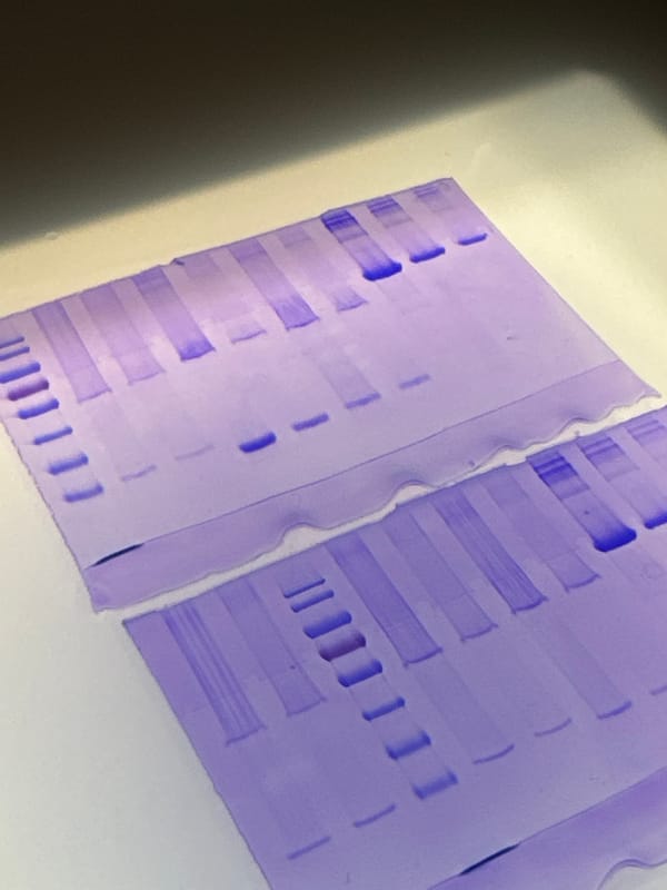
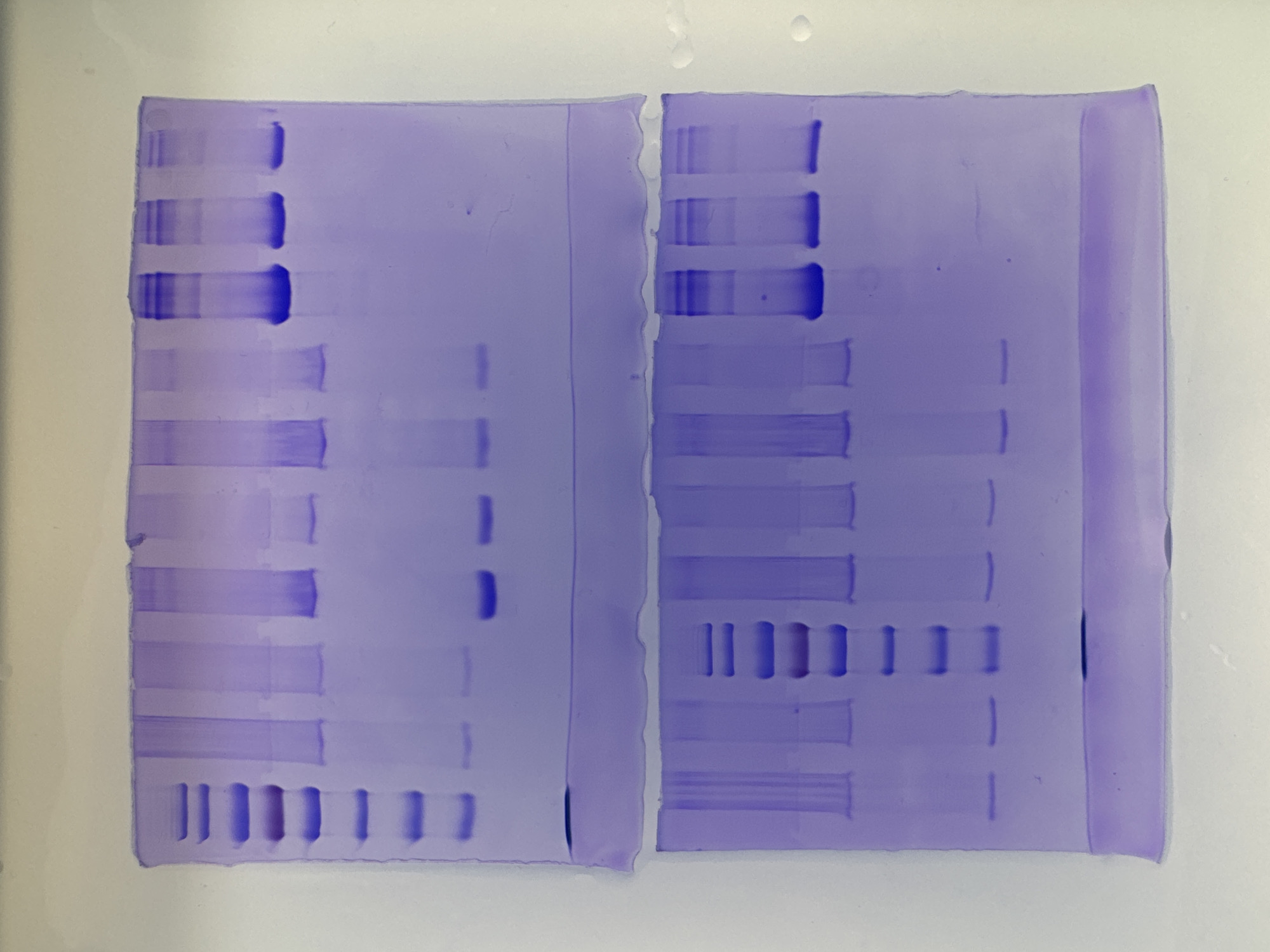
The image displays two purple-stained electrophoretic gels, positioned side-by-side on a light, uniform background. These gels are typical results from a laboratory procedure like SDS-PAGE, used for separating biological molecules such as proteins or DNA. The purple color suggests a common staining method, likely Coomassie Blue. Each gel contains multiple vertical lanes, within which dark violet-blue bands are visible. These bands represent the separated molecules, with their position indicating size or charge, and their intensity correlating with concentration. The left gel features prominent, thick bands in its upper section, with some appearing sharp and others slightly smeared. As one moves down this gel, individual, distinct bands of varying thickness are seen. Towards the bottom, there is a clear pattern of short, uniformly spaced horizontal bands spanning several lanes, characteristic of a molecular weight ladder or control samples. The right gel exhibits similar characteristics. Its upper region also shows several intensely stained bands, some well-defined and others with a degree of smearing. The middle section of this gel conspicuously displays a series of short, distinct bands arranged horizontally across multiple lanes, mirroring the pattern observed at the bottom of the left gel. The lower part of the right gel also contains various horizontal bands of different thicknesses and intensities. The overall image provides a clear view of the stained gels, highlighting the successful separation of samples. There are no people, visible text, or specific location identifiers within the image itself. The lighting is even, suggesting a controlled indoor environment.
Loading AttnAds…
No transactions found