
Sign in
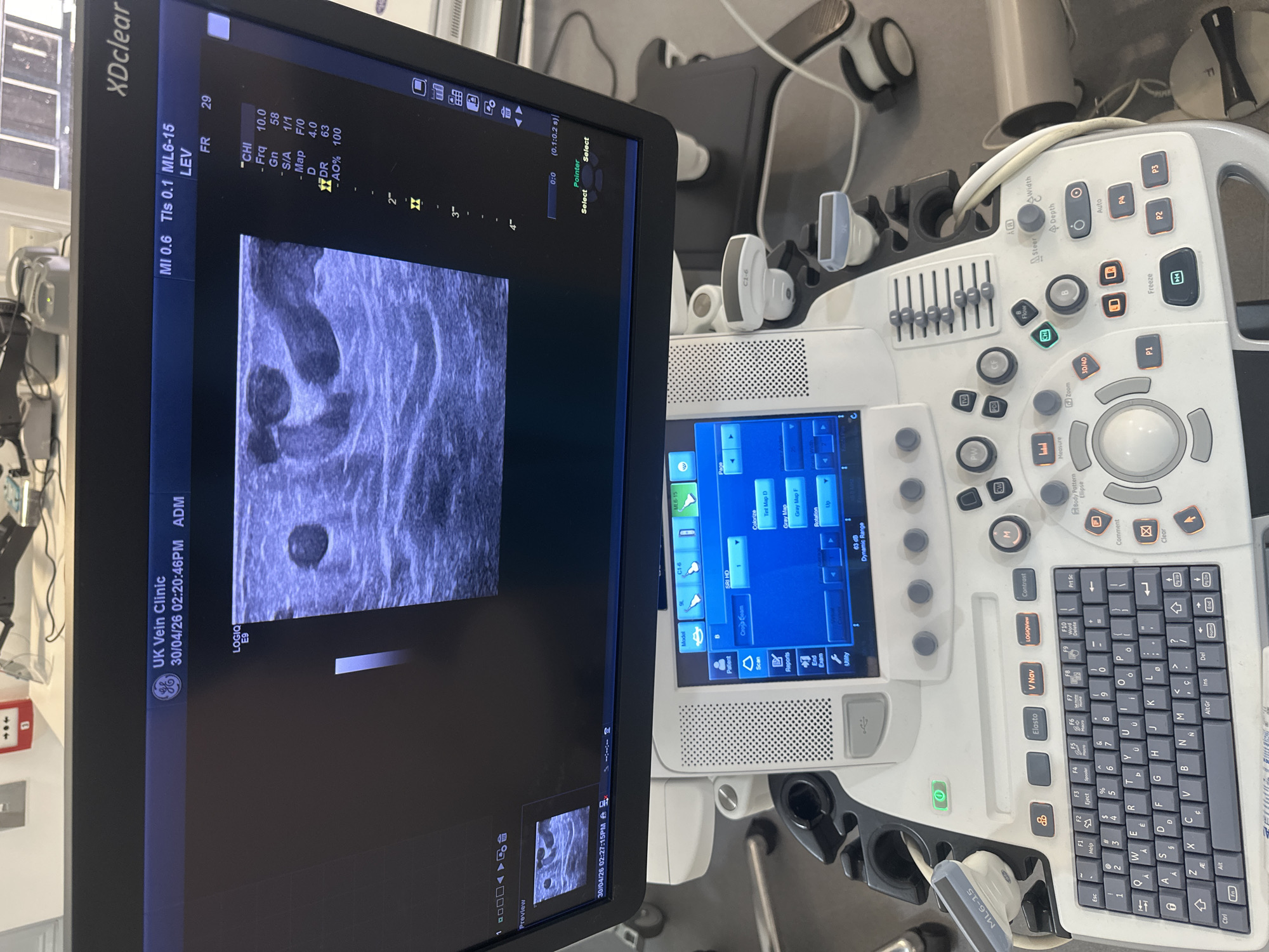
The image displays an ultrasound machine with its monitor and control panel, situated in what appears to be a medical examination room or clinic. The large monitor, branded "XDclear," shows a greyscale ultrasound image with cross-sections of what look like blood vessels, indicative of a vascular scan. Text on the screen identifies the machine as a "GE LOGIQ E9" and the facility as "UK Vein Clinic," confirming a medical, specifically phlebology, setting.

The monitor also displays technical parameters such as "MI 0.6," "Tls 0.1," "ML6-15," "FR 29," "Frq 10.0," "Gn 58," "D 4.0," and "DR 63," which are typical ultrasound settings. The date and time "30/04/26 02:20:46PM" are visible, suggesting an afternoon examination. Below the main image, a smaller preview section shows another ultrasound image with the timestamp "30/04/26 02:27:15PM."

The control panel of the ultrasound machine is visible on the right, featuring a keyboard, various buttons for functions like "Freeze," "Auto," "Depth," "Zoom," and different imaging modes (B-mode, M-mode, PW, CW, CF). Several ultrasound probes, including one labeled "ML6-15," are resting in their holders on the machine. The overall scene suggests an active diagnostic session or recent use of the equipment for vascular assessment in a clinic located in the UK.
kittyorkatherine

Apr 30, 2026, 1:21 PM

London, UK

Moment

Stake attention in this memory

clinical
medical
diagnostic
examination
technology

The image displays an ultrasound machine with its monitor and control panel, situated in what appears to be a medical examination room or clinic. The large monitor, branded "XDclear," shows a greyscale ultrasound image with cross-sections of what look like blood vessels, indicative of a vascular scan. Text on the screen identifies the machine as a "GE LOGIQ E9" and the facility as "UK Vein Clinic," confirming a medical, specifically phlebology, setting. The monitor also displays technical parameters such as "MI 0.6," "Tls 0.1," "ML6-15," "FR 29," "Frq 10.0," "Gn 58," "D 4.0," and "DR 63," which are typical ultrasound settings. The date and time "30/04/26 02:20:46PM" are visible, suggesting an afternoon examination. Below the main image, a smaller preview section shows another ultrasound image with the timestamp "30/04/26 02:27:15PM." The control panel of the ultrasound machine is visible on the right, featuring a keyboard, various buttons for functions like "Freeze," "Auto," "Depth," "Zoom," and different imaging modes (B-mode, M-mode, PW, CW, CF). Several ultrasound probes, including one labeled "ML6-15," are resting in their holders on the machine. The overall scene suggests an active diagnostic session or recent use of the equipment for vascular assessment in a clinic located in the UK.

Symbol

2A4F4

Volume

10,861

Creator

+$0.04

Revenue

+$0.11

TVL

$2.55

1D
1W
1M
All

Loading AttnAds…

transactions
revenues
stakers
Earliest
Latest
Highest stake

1

@anyone

Staked 2mo ago

$2.47 USD

+$0.09 USD

+4%

2

Rev Bot 🤖💰

Injected revenue 2mo ago

Revenue bonus for the last stake.

+$0.12 USD

More from this user

More from London

kittyorkatherine

Apr 30, 2026, 1:21 PM

London, UK

Licensed under Creative Commons Attribution 4.0 International License. You are free to share and adapt this content with proper attribution.