
Stake attention in this memory
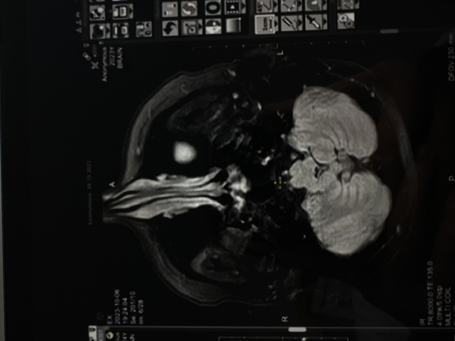
An axial T2-weighted MRI scan of a human brain is displayed on a digital monitor. The grayscale image shows internal cranial structures, with the cerebellum prominent in the inferior portion and a bright, ovoid structure located in the right frontal region of the scan. On the top left, the date "2023-10-06" and time "19:24:04" are visible, along with "Se 201/10" and "Im 6/28", indicating sequence and slice information. The patient is identified as "Anonymous" (top center and right), and the scan type is "BRAIN". Technical parameters "TR 8000.0 TE 135.0", "4 Oth/s 0sp", and "MULTI COIL" are present at the bottom left, with "DFOV 230 mm" at the bottom right. A vertical toolbar on the right side of the screen displays various graphical interface icons, including a "1cm" scale. The image does not contain information identifying a specific location within the city of Kazan, Russia.
Loading AttnAds…
No transactions found