
Sign in
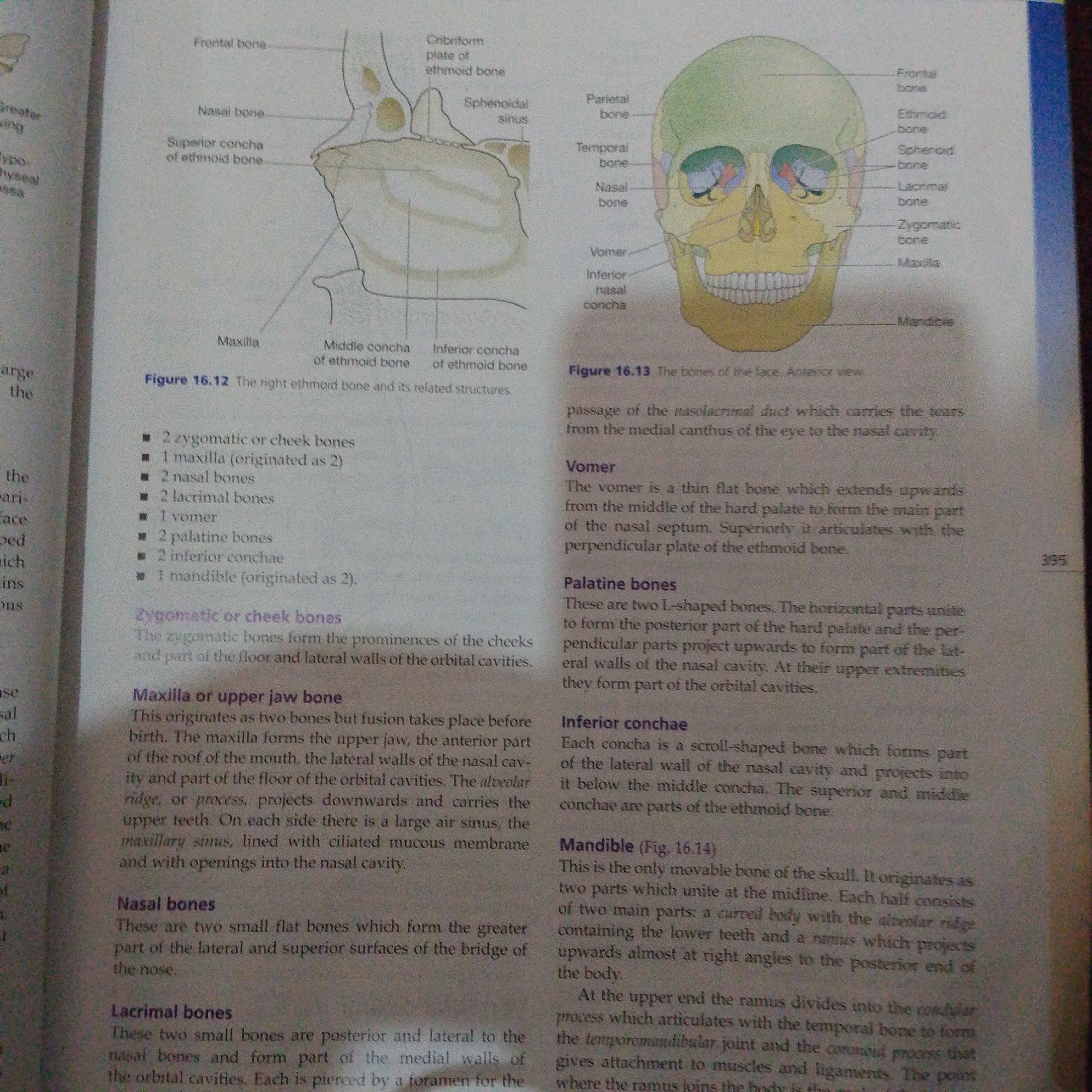
The image shows two anatomical illustrations of the human skull, focusing on the bones of the face and nasal cavity. The left illustration, labeled "Figure 16.12," depicts the ethmoid bone and its related structures, with labels pointing to the frontal bone, nasal bone, superior concha of the ethmoid bone, maxilla, middle concha of the ethmoid bone, and inferior concha of the ethmoid bone. The right illustration, labeled "Figure 16.13," presents an anterior view of the skull, labeling various bones including the frontal bone, parietal bone, temporal bone, sphenoid bone, ethmoid bone, lacrimal bone, zygomatic bone, maxilla, and mandible.

Below the illustrations, there is a list of bones of the face, followed by descriptive text about the zygomatic or cheek bones, maxilla or upper jaw bone, nasal bones, lacrimal bones, vomer, palatine bones, inferior conchae, and the mandible. The text provides details about the origin, location, and function of these bones. For instance, it mentions that the maxilla forms the upper jaw and part of the roof of the mouth, and the mandible is the only movable bone of the skull.

Visible text includes labels for anatomical features and bone names, figure numbers (16.12 and 16.13), and descriptive paragraphs. There are no people, animals, or specific scenes depicted. The setting is a page from an anatomy or biology textbook. The time of day, weather, and location cues are not present in the image. The overall tone is informative and educational.
FM-66btC3

Jun 30, 2026, 1:57 PM

Jalingo, Nigeria

Stake attention in this memory

curiosity
analytical
studious
calm
neutral

The image shows two anatomical illustrations of the human skull, focusing on the bones of the face and nasal cavity. The left illustration, labeled "Figure 16.12," depicts the ethmoid bone and its related structures, with labels pointing to the frontal bone, nasal bone, superior concha of the ethmoid bone, maxilla, middle concha of the ethmoid bone, and inferior concha of the ethmoid bone. The right illustration, labeled "Figure 16.13," presents an anterior view of the skull, labeling various bones including the frontal bone, parietal bone, temporal bone, sphenoid bone, ethmoid bone, lacrimal bone, zygomatic bone, maxilla, and mandible. Below the illustrations, there is a list of bones of the face, followed by descriptive text about the zygomatic or cheek bones, maxilla or upper jaw bone, nasal bones, lacrimal bones, vomer, palatine bones, inferior conchae, and the mandible. The text provides details about the origin, location, and function of these bones. For instance, it mentions that the maxilla forms the upper jaw and part of the roof of the mouth, and the mandible is the only movable bone of the skull. Visible text includes labels for anatomical features and bone names, figure numbers (16.12 and 16.13), and descriptive paragraphs. There are no people, animals, or specific scenes depicted. The setting is a page from an anatomy or biology textbook. The time of day, weather, and location cues are not present in the image. The overall tone is informative and educational.

Loading AttnAds…

transactions
revenues
stakers
Earliest
Latest
Highest stake

No transactions found

More from this user

More from Jalingo

FM-66btC3

Jun 30, 2026, 1:57 PM

Jalingo, Nigeria

Licensed under Creative Commons Attribution 4.0 International License. You are free to share and adapt this content with proper attribution.