
Stake attention in this memory
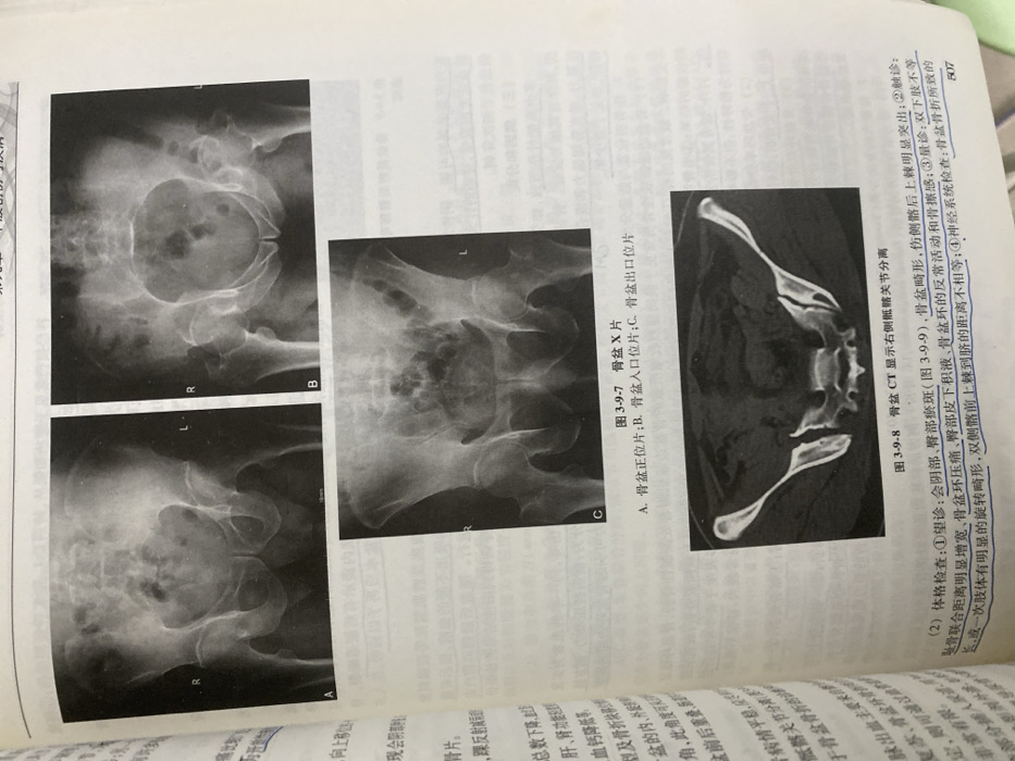
The image shows radiographic images of the pelvis. Figure 3-9-7 presents X-ray images: A is an AP view of the pelvis, B is an inlet view, and C is an outlet view. Figure 3-9-8 shows a CT scan demonstrating right sacroiliac joint separation. Text below outlines physical examination findings, including perineal and gluteal ecchymosis, pelvic deformity, and a prominent posterior superior iliac spine on the injured side. Palpation reveals increased pubic symphysis width, pelvic ring tenderness, and subcutaneous fluid in the buttocks. Limb length inequality and rotational deformity may be present. Neurological examination is indicated to assess for nerve injury related to pelvic fractures.
Loading AttnAds…
No transactions found