Stake attention in this memory
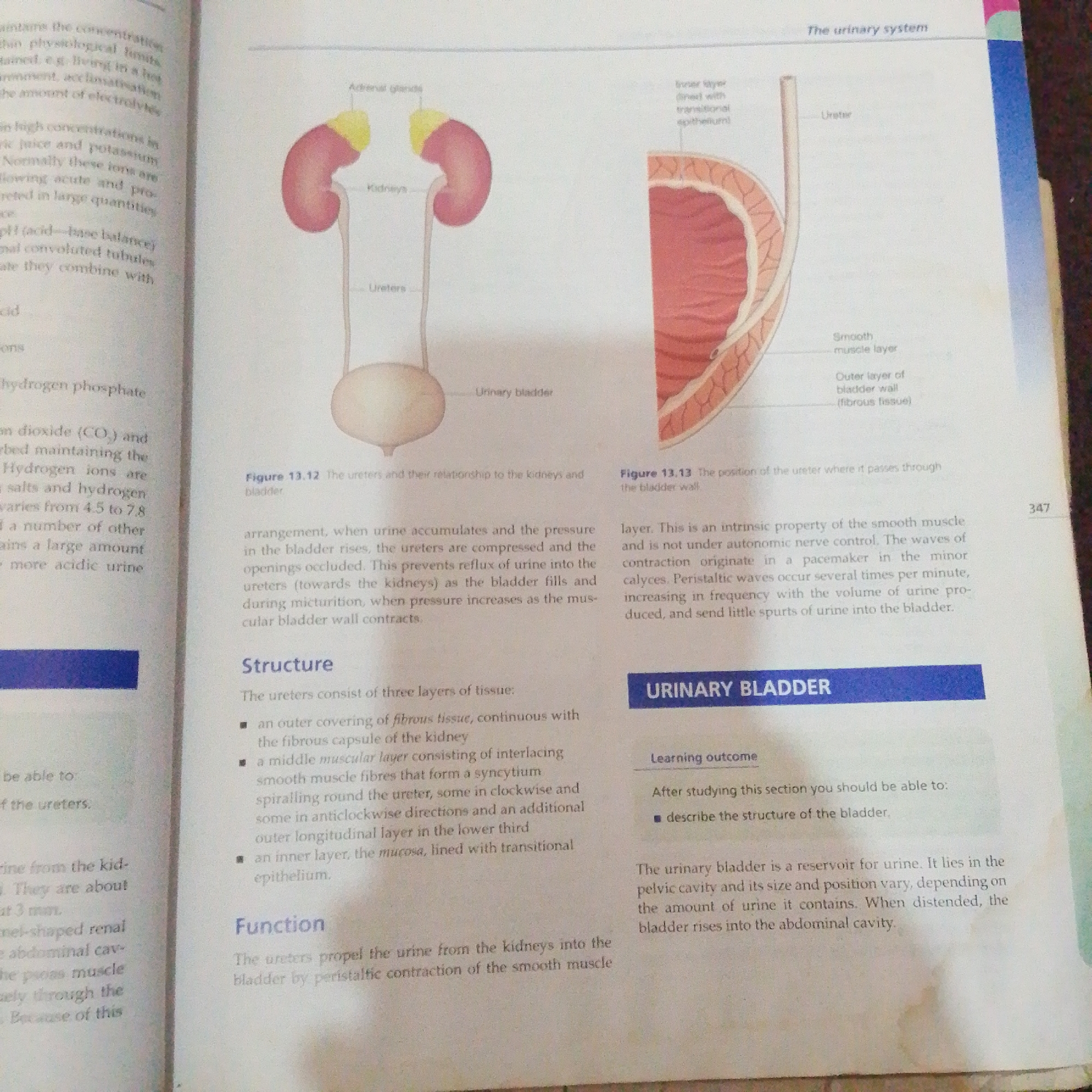
The image displays an illustration of the human urinary system and a cross-section of the bladder wall. On the left, a diagram shows two kidneys with adrenal glands on top, connected by ureters to a urinary bladder. Labels identify these organs. To the right, a detailed cross-section of the bladder wall is shown, with labels indicating the inner layer with transitional epithelium, smooth muscle layer, and outer layer of bladder wall (fibrous tissue). Text boxes on the left discuss physiological limits, hydrogen ions, and pH, while text on the right explains the function of peristaltic waves in urine transport and introduces the urinary bladder as a reservoir. A page number "347" is visible in the top right corner. The image is a textbook illustration and does not depict a real-world scene in Jalingo, Nigeria, or any other location.
No transactions found