
Sign in
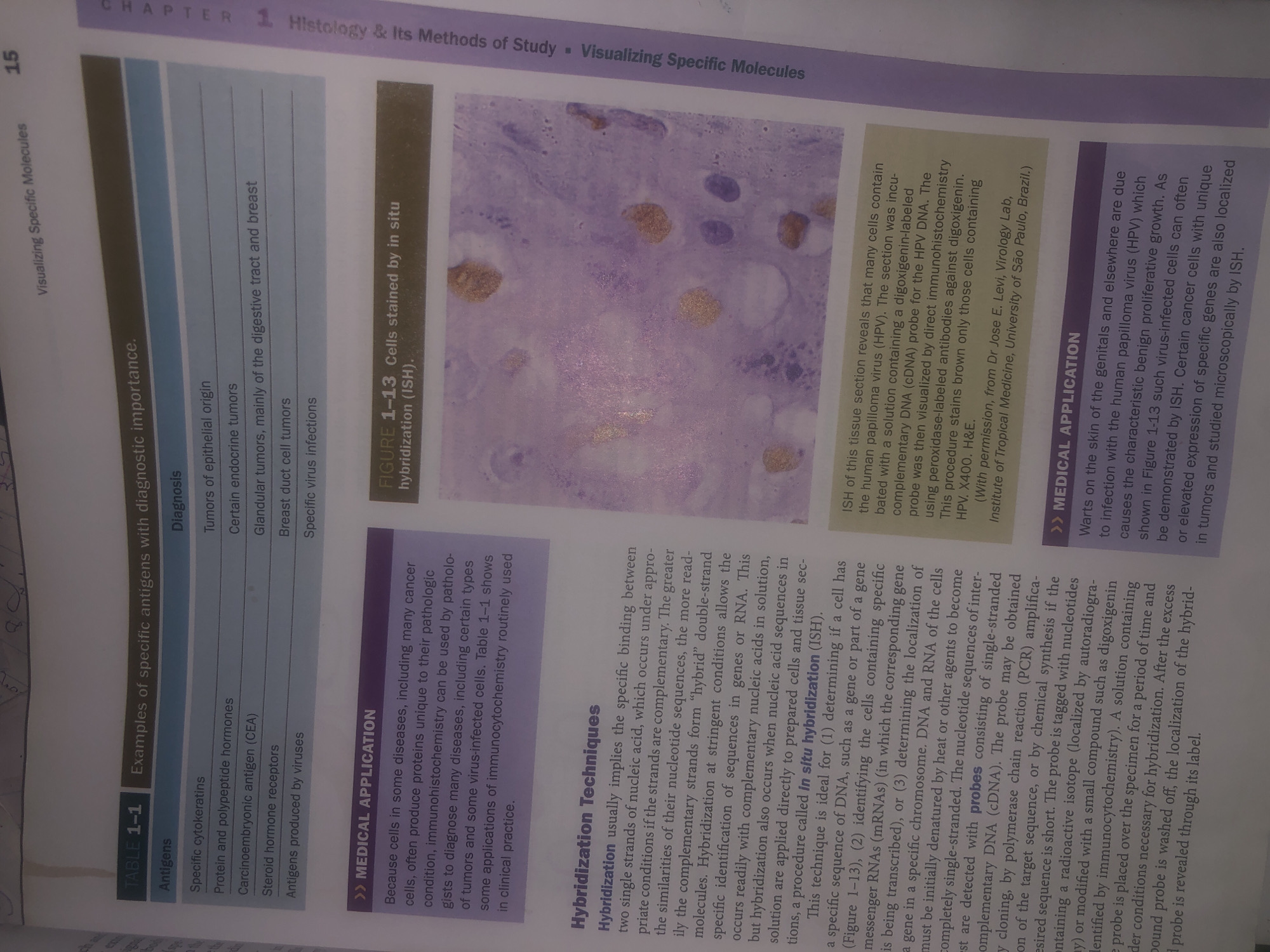
The image displays a page from a textbook or academic publication. It contains text and a micrograph.

The text is divided into sections:
*   **TABLE 1-1 Examples of specific antigens with diagnostic importance:** This table lists various antigens and their associated diagnoses.
*   **>> MEDICAL APPLICATION:** This section discusses the use of immunohistochemistry and immunocytochemistry in diagnosing diseases, particularly cancer and virus-infected cells.
*   **Hybridization Techniques:** This section explains the process of hybridization, particularly in situ hybridization (ISH), a technique used to identify specific nucleic acid sequences.
*   **FIGURE 1-13 Cells stained by in situ hybridization (ISH):** This figure is accompanied by a caption describing a tissue section stained for human papilloma virus (HPV) using ISH, showing virus-containing cells.

The micrograph is a microscopic image showing cells with stained nuclei and cytoplasm. Some cells exhibit brown granular staining, indicating the presence of HPV as described in the caption. The image is labeled with magnification (x400) and a citation.

The provided context does not contain any information that would allow for a description of the location in Wukari, Nigeria.
Bigstev

Jun 12, 2026, 1:34 AM

Wukari, Nigeria

Moment

Stake attention in this memory

informative
scientific
educational
detailed
technical

The image displays a page from a textbook or academic publication. It contains text and a micrograph. The text is divided into sections: * **TABLE 1-1 Examples of specific antigens with diagnostic importance:** This table lists various antigens and their associated diagnoses. * **>> MEDICAL APPLICATION:** This section discusses the use of immunohistochemistry and immunocytochemistry in diagnosing diseases, particularly cancer and virus-infected cells. * **Hybridization Techniques:** This section explains the process of hybridization, particularly in situ hybridization (ISH), a technique used to identify specific nucleic acid sequences. * **FIGURE 1-13 Cells stained by in situ hybridization (ISH):** This figure is accompanied by a caption describing a tissue section stained for human papilloma virus (HPV) using ISH, showing virus-containing cells. The micrograph is a microscopic image showing cells with stained nuclei and cytoplasm. Some cells exhibit brown granular staining, indicating the presence of HPV as described in the caption. The image is labeled with magnification (x400) and a citation. The provided context does not contain any information that would allow for a description of the location in Wukari, Nigeria.

Symbol

08054

Volume

617

Creator

+$0.03

Revenue

+$0.00

TVL

$0.21

1D
1W
1M
All

Loading AttnAds…

transactions
revenues
stakers
Earliest
Latest
Highest stake

1

easydmc

Staked 29d ago
Redeemed 21d ago

$2.17 USD

+$0.00 USD

Fee: $0.01 (0.25%)

+0.0%

2

Rev Bot 🤖💰

Injected revenue 29d ago

Revenue bonus for the last stake.

+$0.03 USD

3

CM🤖

Staked 29d ago

$0.21 USD

-$0.00 USD

-0.2%

More from this user

More from Wukari

Bigstev

Jun 12, 2026, 1:34 AM

Wukari, Nigeria

Licensed under Creative Commons Attribution 4.0 International License. You are free to share and adapt this content with proper attribution.