
Stake attention in this memory
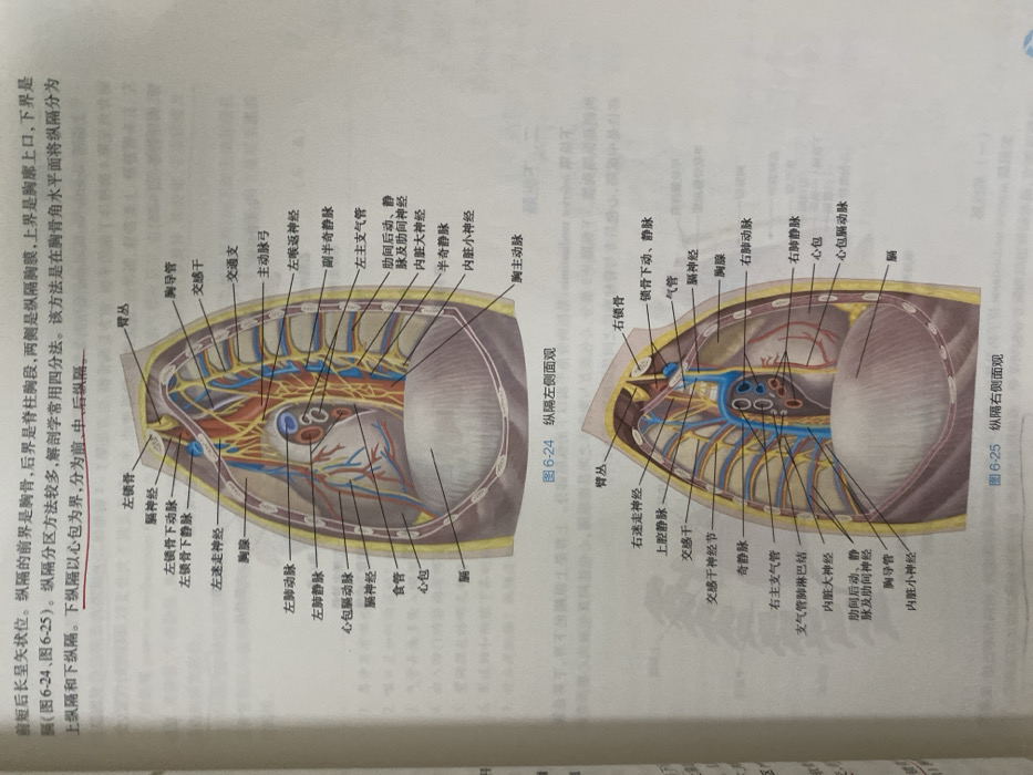
The image displays anatomical illustrations of the mediastinum in the human body. Figure 6-24 showcases a left lateral view, highlighting structures like the left subclavian vessels, phrenic and vagus nerves, aorta, esophagus, and pericardium. Figure 6-25 presents a right lateral view, depicting the right vagus nerve, superior vena cava, sympathetic trunk, azygos vein, trachea, and pericardium. The accompanying text defines the mediastinum and its subdivisions, explaining that it is divided into superior and inferior regions by a plane at the sternal angle and that the inferior region is further divided into anterior, middle and posterior parts with the pericardium as the boundary.
Loading AttnAds…
No transactions found