Stake attention in this memory
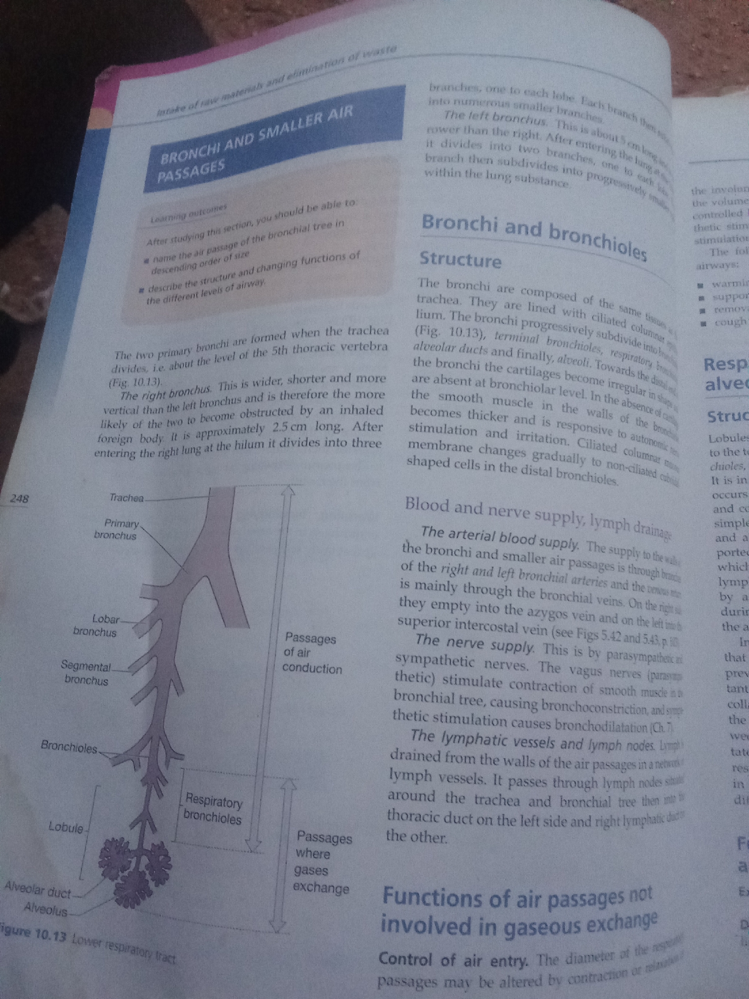
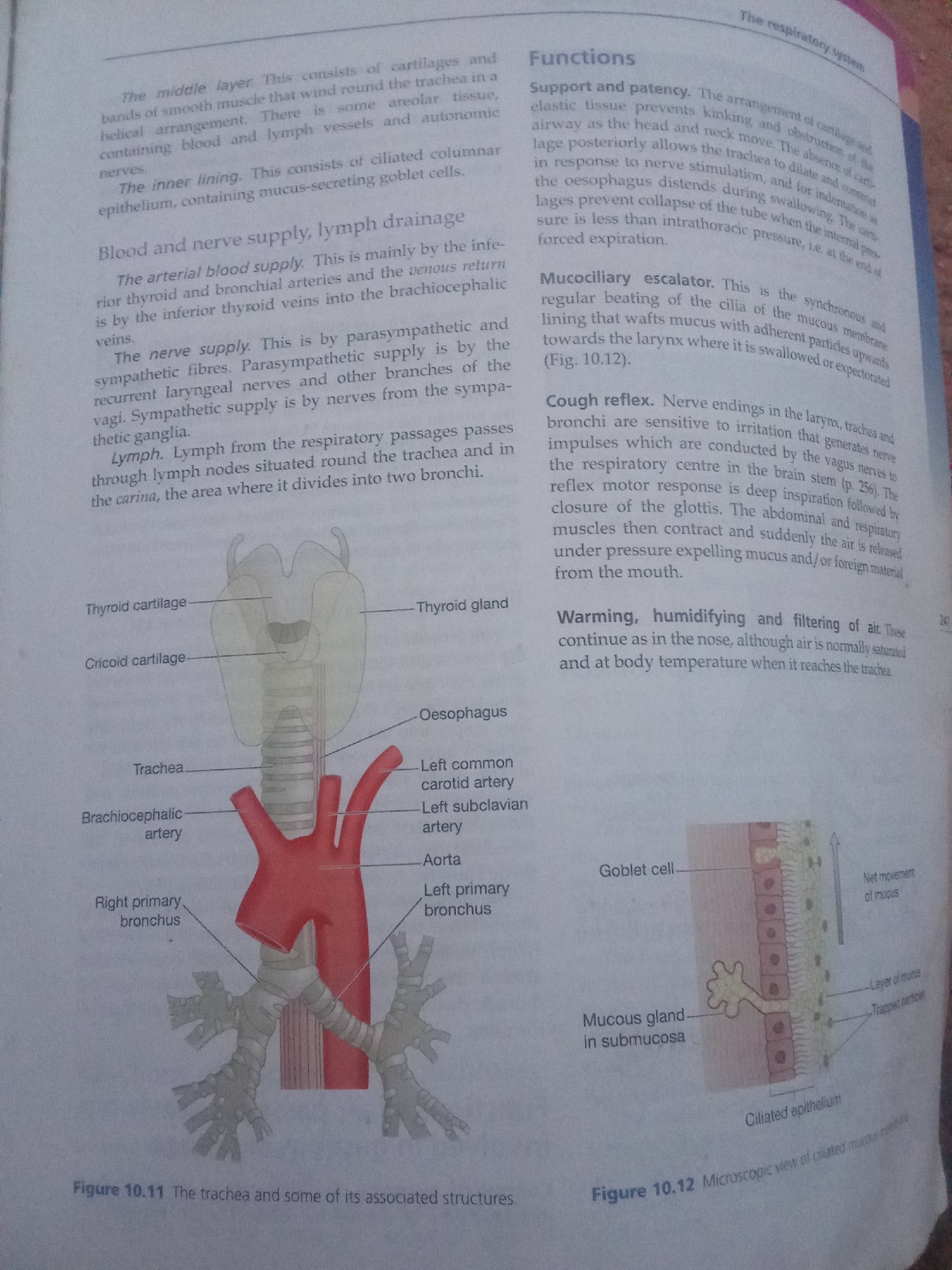
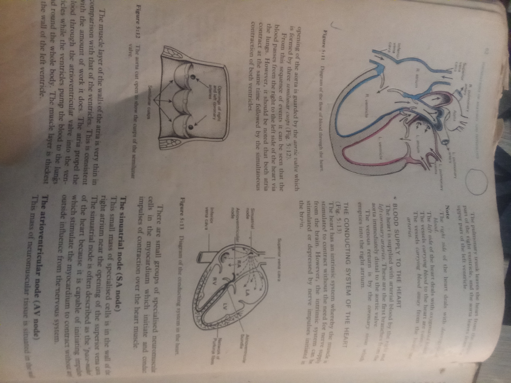
This media file contains scanned pages from a textbook or educational material. The primary subject matter is the anatomy and physiology of the human heart. The pages feature several diagrams illustrating the heart's structure, blood flow, and electrical conduction system. Specifically, there are figures labeled "Figure 5.11 Diagram of the flow of blood through the heart," "Figure 5.12 The aorta cut open to show the cusps of the semilunar valve," and "Figure 5.13 Diagram of the conducting system in the heart." These diagrams are accompanied by detailed text explaining various parts of the heart, such as the atria, ventricles, valves (aortic, atrioventricular), major blood vessels (aorta, pulmonary artery, vena cava), and the sinoatrial (SA) node and atrioventricular (AV) node, which are key components of the heart's conducting system. The text discusses the differences in muscle thickness between the atria and ventricles, relating it to their respective workloads. It also explains the intrinsic nature of the heart's stimulation for contraction, the role of the SA node as the "pace-maker," and the blood supply to the heart itself through the coronary arteries. There are no people, explicit scenes, or environmental settings depicted beyond the textbook pages themselves. The context is purely educational, focusing on biological science. The time of day, weather, and any emotional cues are irrelevant to the content. The location context of Jalingo, Nigeria, is not visually represented or suggested by the content of the images. The visible text is exclusively in English and comprises scientific explanations and labels for the diagrams.
Loading AttnAds…
No transactions found