Stake attention in this memory
anatomy
digestive system
diagram
medical
German
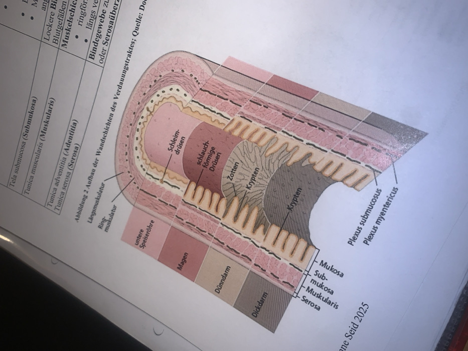
The image depicts a diagram of the layers of the digestive tract wall, labeled in German. Key layers include mucosa, submucosa, muscularis, and serosa (or adventitia). Specific features like "Zotten" (villi), "Krypten" (crypts), "Schleimdrüsen" (mucous glands), and "schlauchförmige Drüsen" (tubular glands) are shown. Muscularis consists of both longitudinal and circular muscle layers. The diagram demonstrates variations in the layer structures across different parts of the digestive system, labeled as "untere Speiseröhre" (lower esophagus), "Magen" (stomach), "Dünndarm" (small intestine), and "Dickdarm" (large intestine). "Plexus submucosus" and "Plexus myentericus" are indicated.
transactions
revenues
stakers
Earliest
Latest
Highest stake
No transactions found