
Stake attention in this memory
radiology
ankylosis
joint dislocation
medical imaging
diagnostic techniques
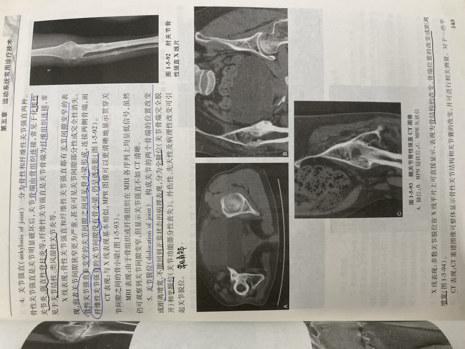
The text describes joint ankylosis and dislocation. Joint ankylosis, either bony or fibrous, involves a narrowed joint space; bony ankylosis may show bone trabeculae bridging the gap, while fibrous ankylosis appears translucent. CT scans better reveal trabeculae than MRI. Joint dislocation, either complete or partial, alters the positions of the articulating bones. X-rays directly show structural and positional changes. CT reconstructions display bony structures and joint capsules for measurement. Images depict elbow bony ankylosis on X-ray (Figure 1-5-92) and hip bony ankylosis on CT scans (Figure 1-5-93) in axial, coronal, and sagittal planes.
Loading AttnAds…
transactions
revenues
stakers
Earliest
Latest
Highest stake
No transactions found