Stake attention in this memory
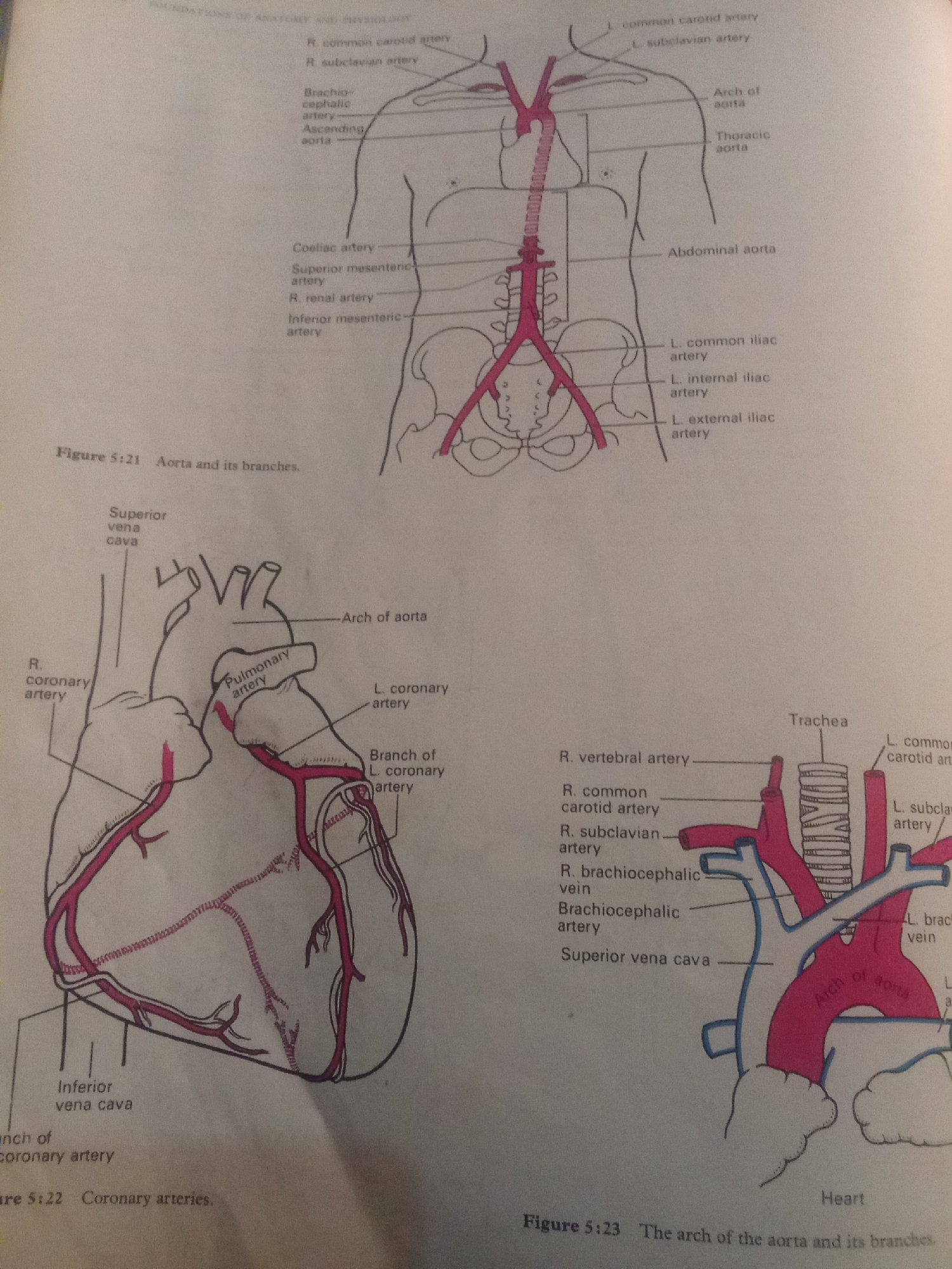
This media file contains three anatomical illustrations related to the circulatory system. The first illustration, labeled "Figure 5:21 Aorta and its branches," depicts the aorta and its major arteries originating from the chest and extending down through the abdomen. Labels identify structures like the common carotid arteries, subclavian arteries, brachiocephalic artery, ascending aorta, arch of the aorta, thoracic aorta, abdominal aorta, and iliac arteries. The second illustration, labeled "Figure 5:22 Coronary arteries," shows a detailed view of the human heart with its coronary arteries. Labels indicate the superior vena cava, pulmonary artery, and the right and left coronary arteries branching across the heart's surface. The third illustration, labeled "Figure 5:23 The arch of the aorta and its branches," focuses on the upper portion of the aorta and the major arteries supplying the head and arms. Labels include the trachea, vertebral artery, common carotid artery, subclavian artery, brachiocephalic vein, brachiocephalic artery, and superior vena cava. The arch of the aorta is prominently featured. All three illustrations are line drawings with red coloring highlighting the arteries. There are no people, scenes, or specific location cues beyond the general anatomical context. The implied setting is likely an educational or medical environment where anatomical diagrams are used for study. There are no indications of time of day, weather, or emotions.
No transactions found