
Stake attention in this memory
histology
submandibular gland
microscopy
stained cells
tissue analysis
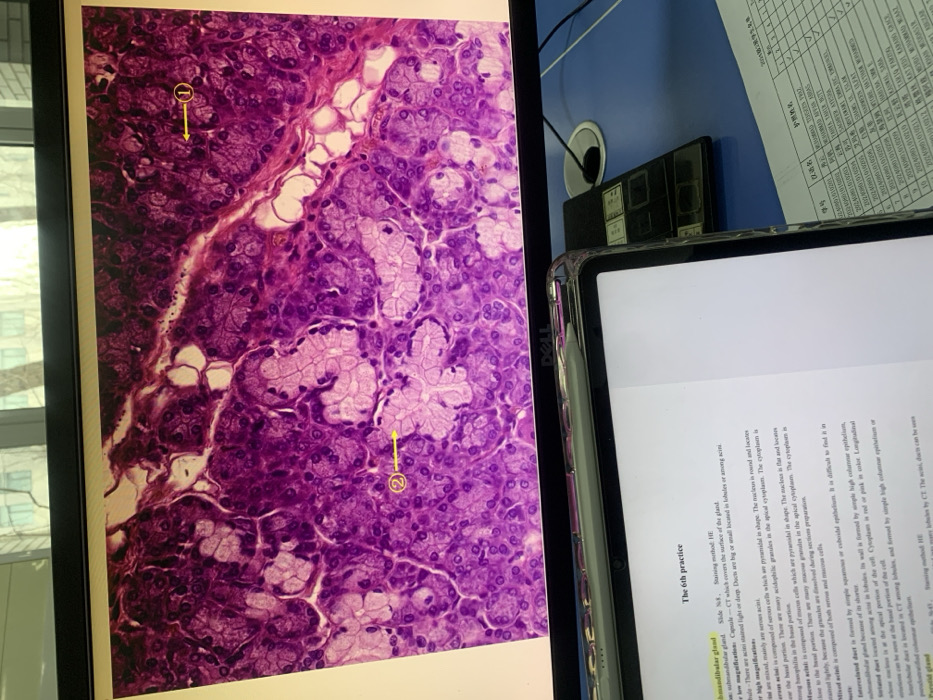
The image shows a histology slide of a submandibular gland stained with Hematoxylin and Eosin (H&E). There are two marked areas on the histological slide. Area 1 shows a connective tissue capsule (CT) which covers the surface of the gland. The slide contains acini which are stained light or dark, ducts of varying sizes in lobules. At higher magnification, it shows mixed acini composed of serous cells which are pyramidal in shape, the nucleus is round in the basal portion, with granules in the apical cytoplasm, and also mucous cells with far nucleus and granules in the apical cytoplasm.
Loading AttnAds…
transactions
revenues
stakers
Earliest
Latest
Highest stake
No transactions found