Stake attention in this memory
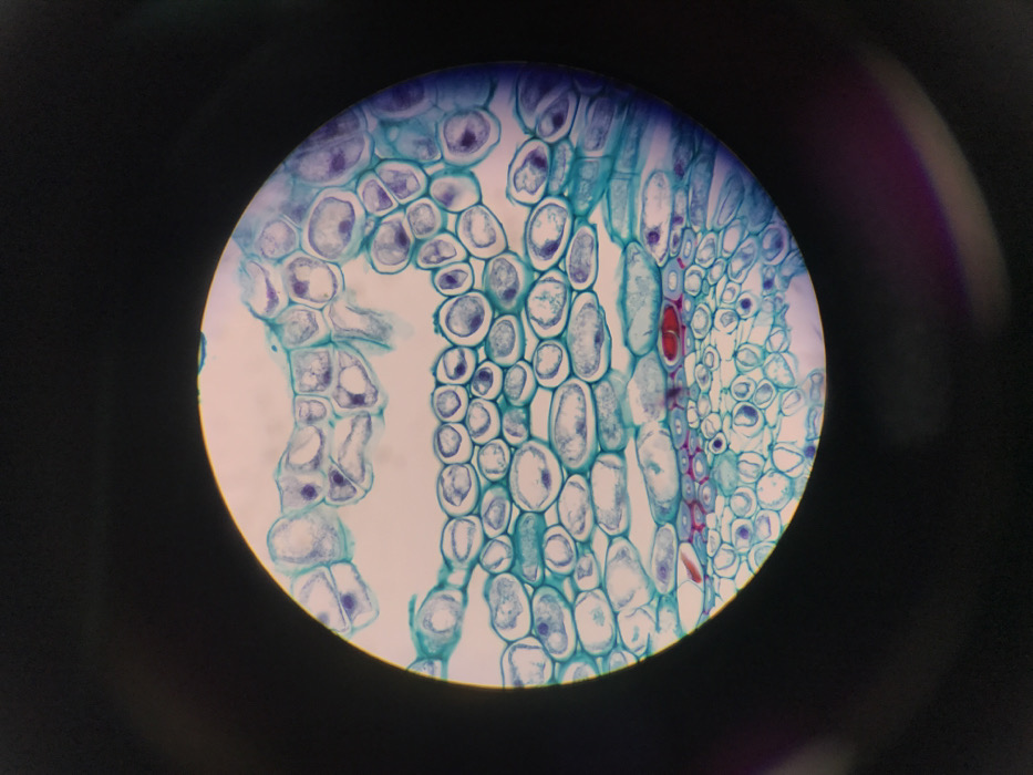
This image presents a circular, highly magnified view of a stained biological tissue section, captured through a light microscope, against a solid black background. The observation took place in Midrand, South Africa. The primary subject is a complex arrangement of biological cells, predominantly stained in shades of blue and purple, with discernible nuclei or dense cytoplasmic components appearing as darker purplish structures within. The cellular architecture displays multiple distinct layers and morphologies. The upper region contains irregularly shaped, polygonal cells. A prominent, horizontally oriented band of uniformly shaped, ovoid to elongated cells, stained bright blue with darker purple interiors, occupies the central portion of the field. Directly below this band, a distinct layer features several cells stained bright red, which are ovoid or irregularly shaped, interspersed among the blue and purple cells. The lowest visible cellular layer consists of smaller, densely packed cells, also stained in blue and purple hues. No individuals are present in the scene. The image records the microscopic structural details of the specimen as illuminated and magnified by a light microscope.
No transactions found