Sign in
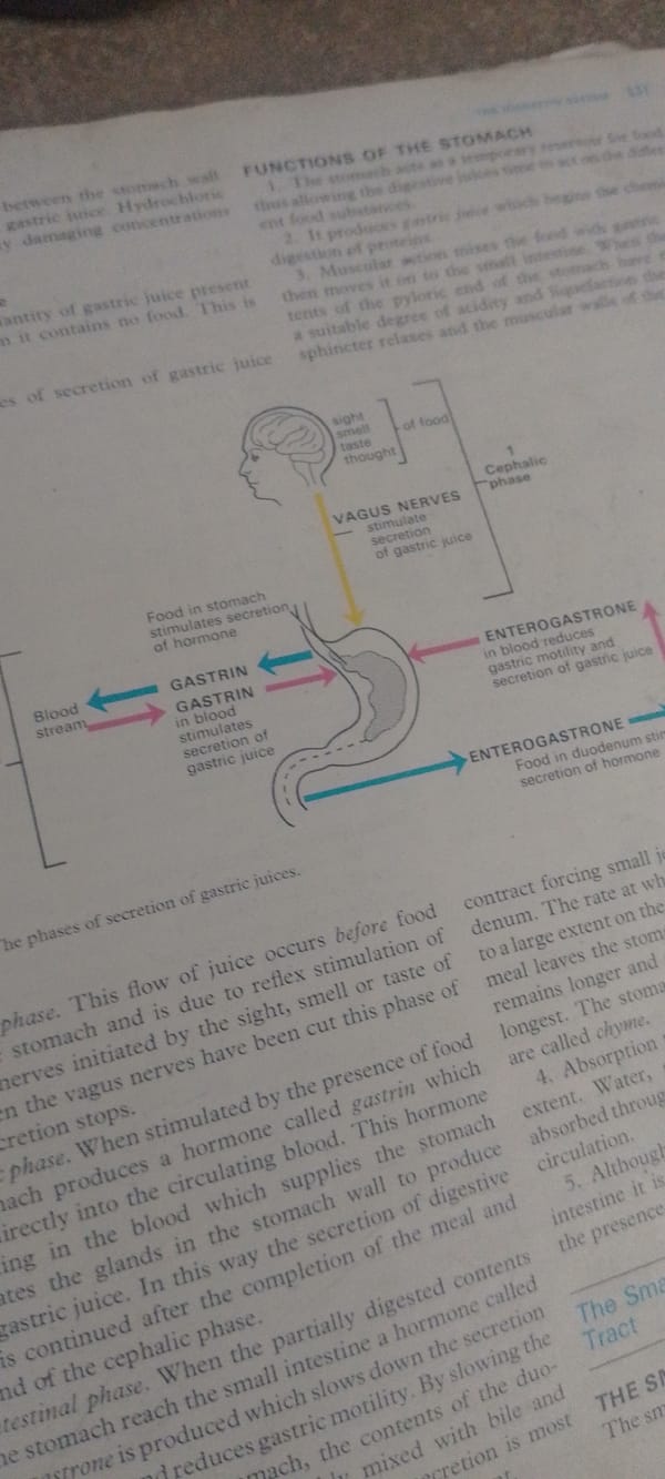
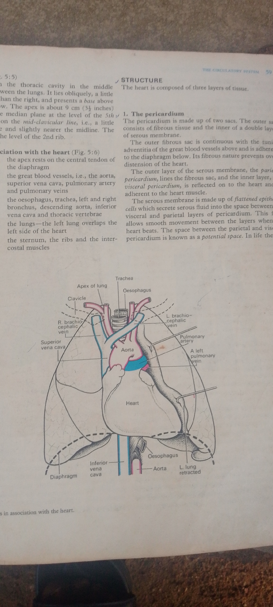
This is a close-up, high-angle shot of an open textbook page, focusing on an anatomical illustration and accompanying text. The subjects are the printed words and diagram, with no people or live elements present. The page is from a book titled "THE CIRCULATORY SYSTEM," as indicated by the header text.

The central focus is a detailed diagram illustrating the human heart and its surrounding structures, including the lungs, trachea, esophagus, aorta, and major blood vessels. Labels in clear, black text identify each part of the illustration, such as "Apex of lung," "Clavicle," "Superior vena cava," "Heart," "Aorta," "Pulmonary artery," and "Diaphragm." Some parts of the diagram are colored, with the heart itself depicted in pink and blue, and certain vessels highlighted in blue and pink as well.

The text discusses the structure of the heart, specifically detailing the pericardium. It describes the two sacs that make up the pericardium, their composition, and their relationship to the heart and surrounding vessels. The text mentions that the pericardium is made up of flattened epithelial cells that secrete serous fluid, allowing for smooth movement of the heart. The page is well-lit, suggesting an indoor setting likely a classroom or study area. There are no discernible emotions or specific time or weather cues. The overall scene is educational and informational, presenting biological knowledge through text and illustration. The bottom of the image shows a dark, textured surface, possibly the cover or a table, beneath the book.
FM-2L6Lq2

Jun 30, 2026, 4:57 PM

Jauro Yinu, Nigeria

Moment

Stake attention in this memory

analytical
intellectual
studious
focused
neutral

This is a close-up, high-angle shot of an open textbook page, focusing on an anatomical illustration and accompanying text. The subjects are the printed words and diagram, with no people or live elements present. The page is from a book titled "THE CIRCULATORY SYSTEM," as indicated by the header text. The central focus is a detailed diagram illustrating the human heart and its surrounding structures, including the lungs, trachea, esophagus, aorta, and major blood vessels. Labels in clear, black text identify each part of the illustration, such as "Apex of lung," "Clavicle," "Superior vena cava," "Heart," "Aorta," "Pulmonary artery," and "Diaphragm." Some parts of the diagram are colored, with the heart itself depicted in pink and blue, and certain vessels highlighted in blue and pink as well. The text discusses the structure of the heart, specifically detailing the pericardium. It describes the two sacs that make up the pericardium, their composition, and their relationship to the heart and surrounding vessels. The text mentions that the pericardium is made up of flattened epithelial cells that secrete serous fluid, allowing for smooth movement of the heart. The page is well-lit, suggesting an indoor setting likely a classroom or study area. There are no discernible emotions or specific time or weather cues. The overall scene is educational and informational, presenting biological knowledge through text and illustration. The bottom of the image shows a dark, textured surface, possibly the cover or a table, beneath the book.

Symbol

1AF46

Volume

10,132

Creator

+$0.03

Revenue

+$0.06

TVL

$2.33

1D
1W
1M
All

Loading AttnAds…

transactions
revenues
stakers
Earliest
Latest
Highest stake

1

FM-FSRgn1

Staked 16d ago

$2.29 USD

+$0.04 USD

+2%

2

Rev Bot 🤖💰

Injected revenue 16d ago

Revenue bonus for the last stake.

+$0.07 USD

More from this user

More from Jauro Yinu

FM-2L6Lq2

Jun 30, 2026, 4:57 PM

Jauro Yinu, Nigeria

Licensed under Creative Commons Attribution 4.0 International License. You are free to share and adapt this content with proper attribution.