Stake attention in this memory
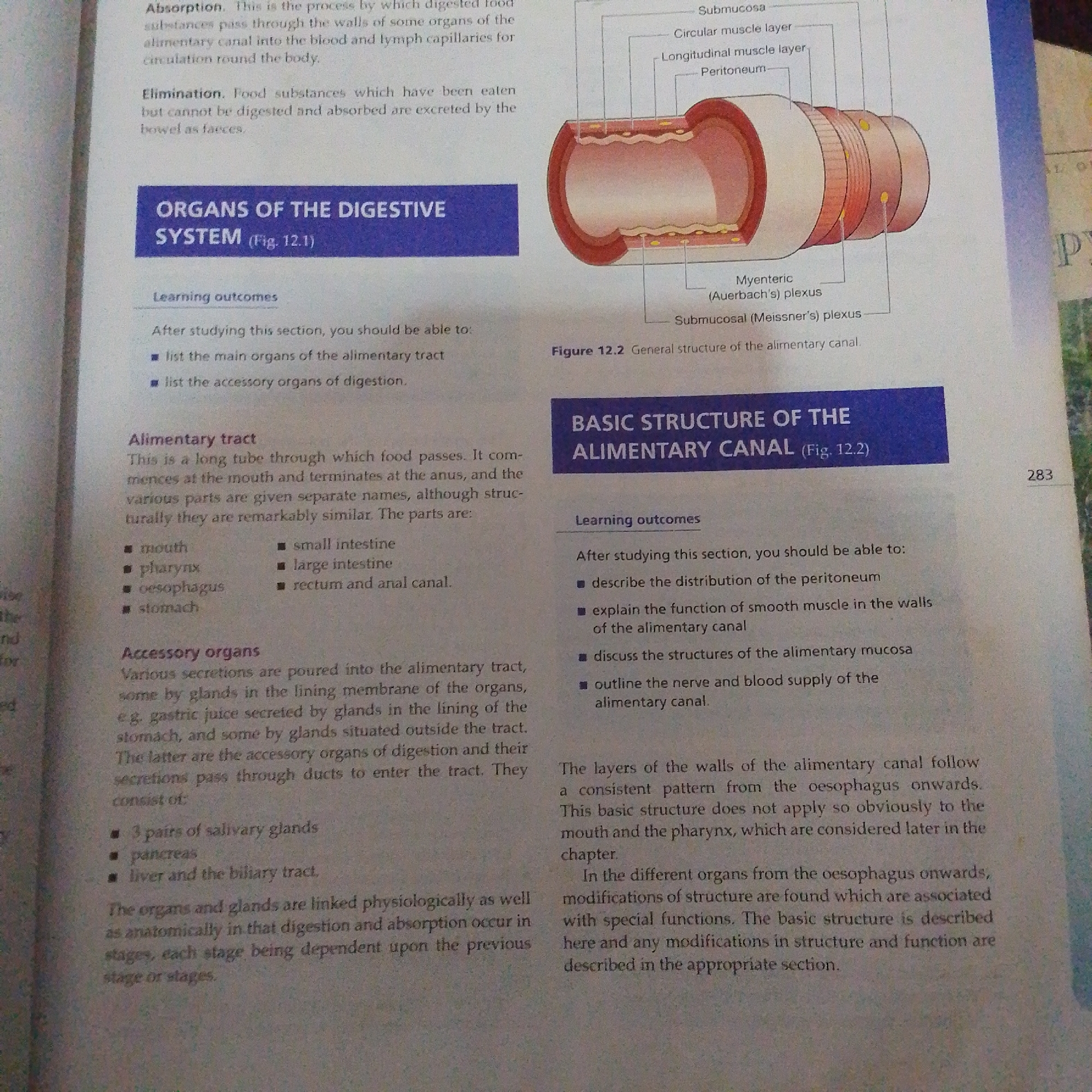
This media file is a scanned page from a textbook, likely a biology or anatomy textbook, discussing the digestive system. The page is divided into two main sections. The left section is titled "ORGANS OF THE DIGESTIVE SYSTEM (Fig. 12.1)" and lists learning outcomes related to identifying main and accessory organs of the alimentary tract. It then details the "Alimentary tract" with a bulleted list of its parts, and "Accessory organs" with a similar list. The text describes the functions of these organs. The right section is titled "BASIC STRUCTURE OF THE ALIMENTARY CANAL (Fig. 12.2)" and also presents learning outcomes, focusing on the peritoneum, smooth muscle function, alimentary mucosa, and nerve/blood supply. This section is accompanied by an illustration of a cross-section of the alimentary canal, showing its various layers: Submucosa, Circular muscle layer, Longitudinal muscle layer, Peritoneum, Myenteric (Auerbach's) plexus, and Submucosal (Meissner's) plexus. The text explains that the basic structure is consistent from the esophagus onwards, with modifications in different organs. A page number "283" is visible in the bottom right corner. The image is well-lit and the text is clear and legible, suggesting it's a high-quality scan. There are no people or specific location cues within the image content itself, other than the general subject matter of human anatomy. The overall scene is that of an academic document.
No transactions found