
Sign in
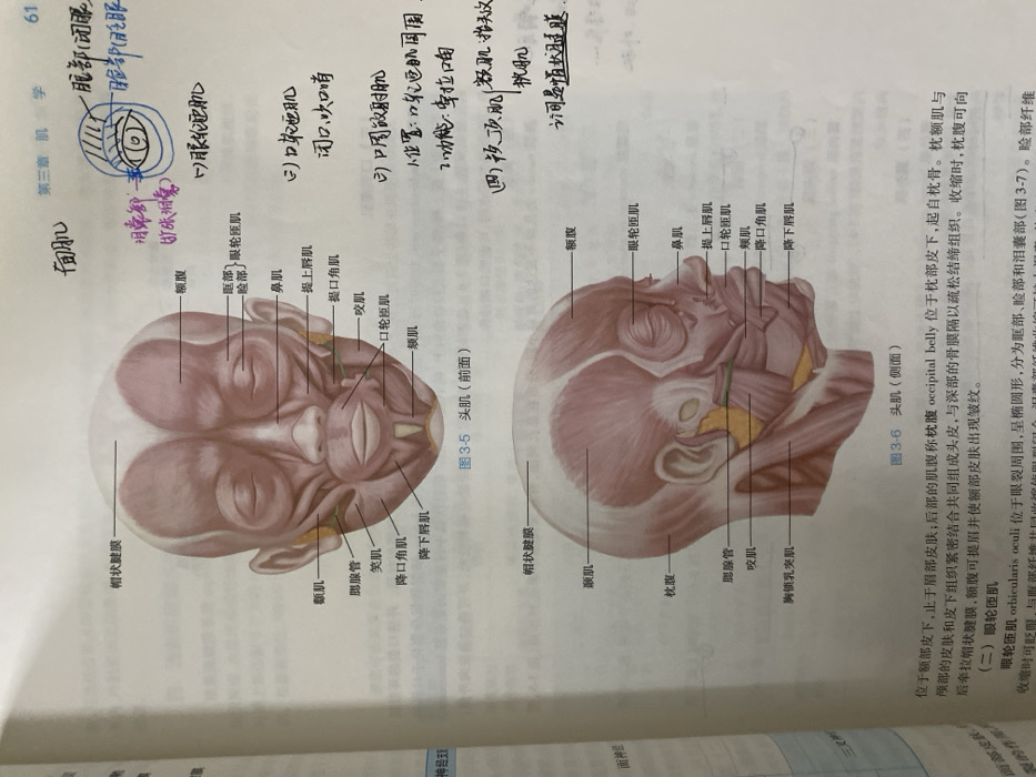
The image presents two anatomical diagrams of the head muscles, titled "Frontal Muscle (Front View)" and "Frontal Muscle (Side View)". Key muscles are labeled, including the epicranial aponeurosis, frontalis, temporalis, orbicularis oculi, orbicularis oris, nasalis, levator labii superioris, levator anguli oris, buccinator, zygomaticus major, depressor anguli oris, depressor labii inferioris, parotid gland, masseter, sternocleidomastoid, and occipitalis. Functional descriptions accompany some muscles, and brief notes describe the occipitofrontalis' location and function.
LANDLORD242

Dec 24, 2025, 4:58 AM

Chongqing High-tech Zone, China

Stake attention in this memory

anatomy
muscles
head
illustration
textbook

The image presents two anatomical diagrams of the head muscles, titled "Frontal Muscle (Front View)" and "Frontal Muscle (Side View)". Key muscles are labeled, including the epicranial aponeurosis, frontalis, temporalis, orbicularis oculi, orbicularis oris, nasalis, levator labii superioris, levator anguli oris, buccinator, zygomaticus major, depressor anguli oris, depressor labii inferioris, parotid gland, masseter, sternocleidomastoid, and occipitalis. Functional descriptions accompany some muscles, and brief notes describe the occipitofrontalis' location and function.

Loading AttnAds…

transactions
revenues
stakers
Earliest
Latest
Highest stake

No transactions found

More from this user

More from Chongqing High-tech Zone

LANDLORD242

Dec 24, 2025, 4:58 AM

Chongqing High-tech Zone, China

Licensed under Creative Commons Attribution 4.0 International License. You are free to share and adapt this content with proper attribution.