Stake attention in this memory
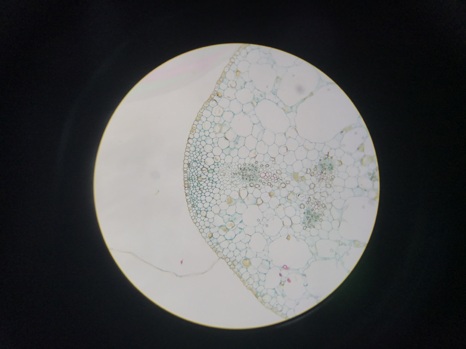
The image presents a bright, circular field of view against a solid black background, characteristic of an optical microscope observation. Within this field, a stained cross-section of plant tissue is visible. The specimen exhibits a curved upper surface composed of tightly packed, small, uniform cells, consistent with an epidermal layer. Beneath this, the tissue transitions to larger, less densely arranged cells identified as parenchyma, some containing small, brownish-yellow granular structures. Deeper within the tissue, several clusters of smaller cells with distinct staining patterns are present, including reddish-pink and light blue elements, indicative of vascular bundles. No people, active subjects, or dynamic interactions are depicted within the frame. This microscopic observation was made in Midrand, South Africa.
No transactions found