Stake attention in this memory
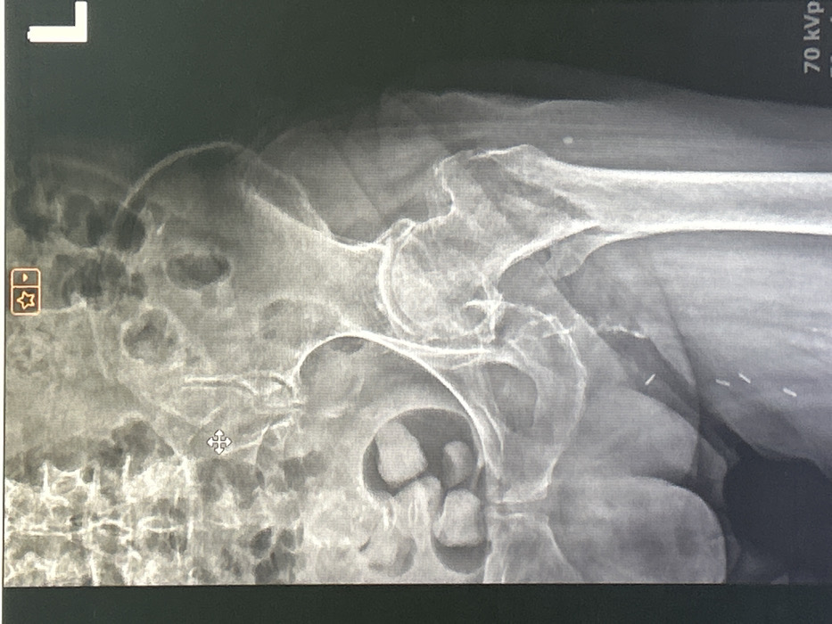
This image is a photograph of a digital anteroposterior radiograph displaying a human pelvis and proximal left femur, originating from the city of Ruse, Bulgaria. The "L" marker in the upper right corner confirms the left anatomical side. The radiograph clearly visualizes the left ilium, ischium, pubis, sacrum, femoral head, neck, and proximal femoral shaft. The left hip joint space appears narrowed with evidence of osteophyte formation, indicative of degenerative changes. Multiple ovoid, radiopaque densities are present in the right lower quadrant of the pelvic cavity, consistent with calcified structures. Additionally, at least three small, linear, radiopaque metallic objects are observed within the soft tissues of the left superior thigh/groin region. Technical exposure parameters, "70 kVp" and "mA," are visible in the bottom right corner. Interface elements such as a crosshair cursor and bookmark icons are present at the top, indicating the image is captured from a digital medical imaging workstation screen. No individuals or direct actions are depicted beyond the displayed medical scan.
Symbol
22B85
Volume
670
Creator
+$0.00
Revenue
+$0.00
TVL
$0.24