Stake attention in this memory
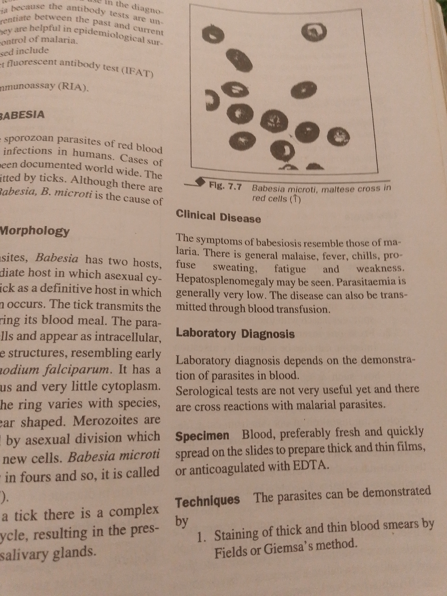
This is a close-up, overhead photograph of an open book. The text on the pages discusses the parasitic disease babesiosis, with specific mention of *Babesia microti*. The left page details the morphology of the parasite, describing its life cycle in ticks and its appearance in red blood cells, noting similarities to *Plasmodium falciparum*. The right page covers clinical disease symptoms, which are similar to malaria, and laboratory diagnosis methods, including microscopic examination of blood smears and serological tests. A scientific illustration in the upper right corner depicts *Babesia microti* in red blood cells, labeled as "Fig. 7.7 Babesia microti, maltese cross in red cells (↑)". The text is black and clearly legible against a white background. The lighting suggests an indoor setting, likely a library or study area.
No transactions found