Stake attention in this memory
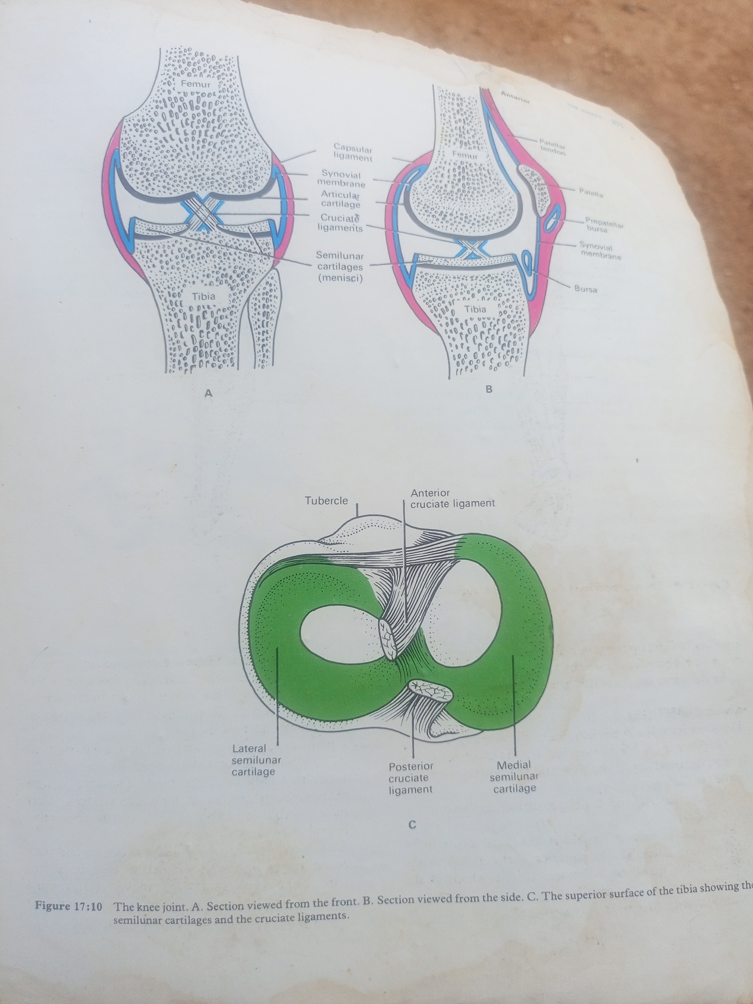
The media file is an illustration of the human knee joint, depicting its anatomical structures. It is not a photograph of a real-world scene or people. The illustration is divided into three parts: A, B, and C. Part A shows a frontal section of the knee joint, labeling structures like the femur, tibia, capsular ligament, synovial membrane, articular cartilage, cruciate ligaments, and semilunar cartilages (menisci). Part B presents a sagittal view of the knee, identifying the patella, patellar tendon, prepatellar bursa, and synovial membrane, along with the femur and tibia. Part C is a view of the superior surface of the tibia, illustrating the semilunar cartilages and cruciate ligaments. The illustration is accompanied by a caption: "Figure 17:10 The knee joint. A. Section viewed from the front. B. Section viewed from the side. C. The superior surface of the tibia showing the semilunar cartilages and the cruciate ligaments." There are no people, animals, or a specific setting depicted. The focus is purely on the scientific and anatomical representation of the knee joint. The image quality suggests it is from a textbook or educational material. The environment where the image was taken appears to be outdoors with natural lighting, as indicated by the shadows and the texture of the surface the material is placed upon, but this context is external to the illustration itself. The location context provided, "Jalingo, Nigeria," does not relate to the content of the image, which is a general anatomical diagram.
No transactions found