
Sign in
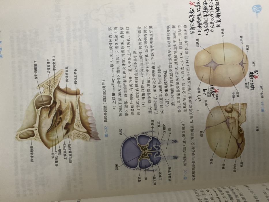
The image shows anatomical illustrations of the human skull, focusing on the nasal cavity and the skull of a newborn. 

Diagram 1-32 depicts the lateral wall of the nasal cavity, with labeled structures like the maxillary sinus, ethmoid bulla, and various sinus openings. Diagram 1-33 is a coronal section of the skull through the third molar, highlighting the oral cavity, palate, and maxillary sinus. Diagram 1-34 illustrates the newborn skull, showing features like fontanelles and ossification centers, along with labels of bones like the frontal, parietal, and occipital bones. The text describes the characteristics of these structures and their clinical significance.
LANDLORD242

Dec 24, 2025, 5:06 AM

Chongqing High-tech Zone, China

Stake attention in this memory

anatomy
skull
sinuses
illustration
textbook

The image shows anatomical illustrations of the human skull, focusing on the nasal cavity and the skull of a newborn. Diagram 1-32 depicts the lateral wall of the nasal cavity, with labeled structures like the maxillary sinus, ethmoid bulla, and various sinus openings. Diagram 1-33 is a coronal section of the skull through the third molar, highlighting the oral cavity, palate, and maxillary sinus. Diagram 1-34 illustrates the newborn skull, showing features like fontanelles and ossification centers, along with labels of bones like the frontal, parietal, and occipital bones. The text describes the characteristics of these structures and their clinical significance.

Loading AttnAds…

transactions
revenues
stakers
Earliest
Latest
Highest stake

No transactions found

More from this user

More from Chongqing High-tech Zone

LANDLORD242

Dec 24, 2025, 5:06 AM

Chongqing High-tech Zone, China

Licensed under Creative Commons Attribution 4.0 International License. You are free to share and adapt this content with proper attribution.