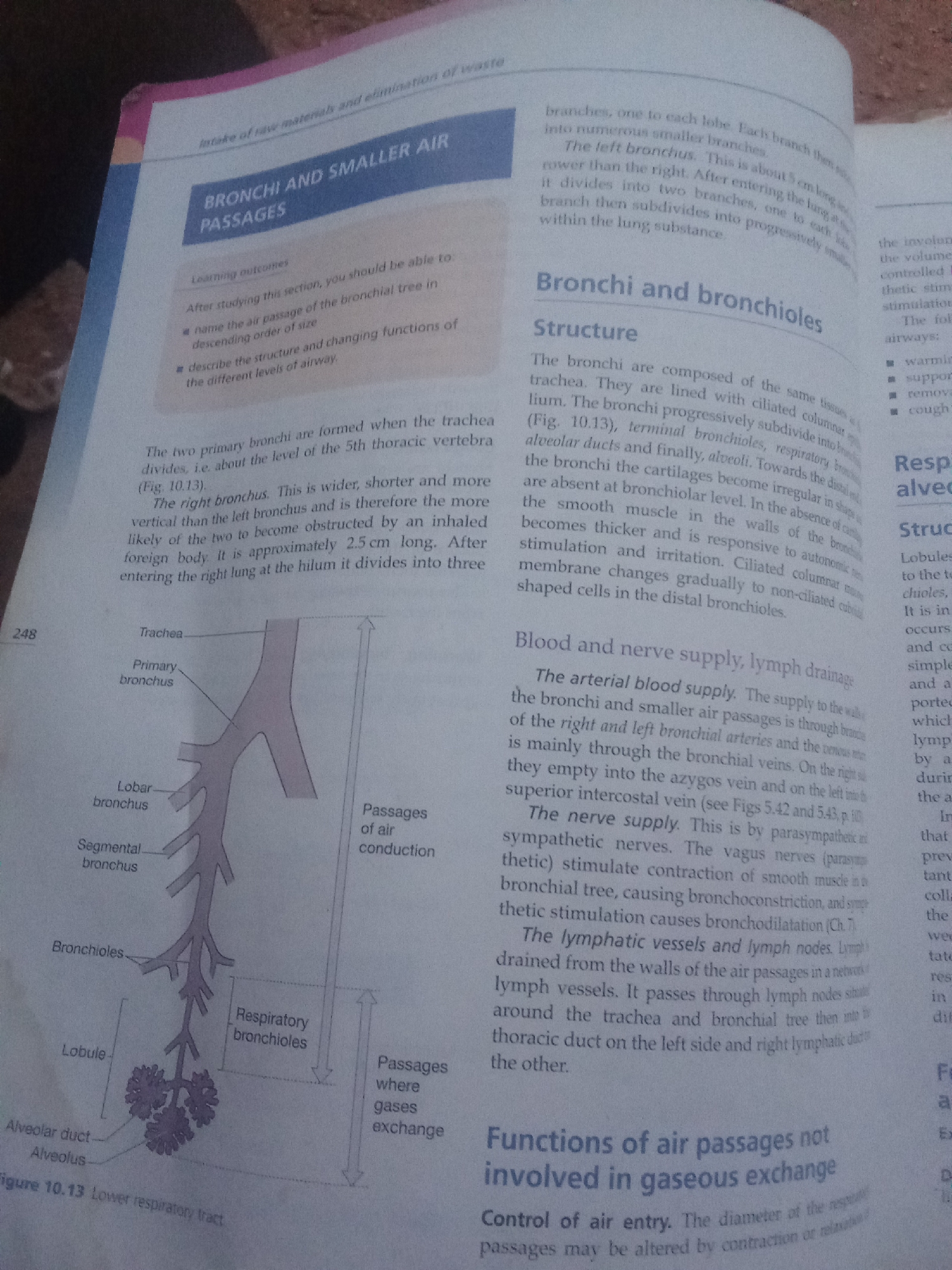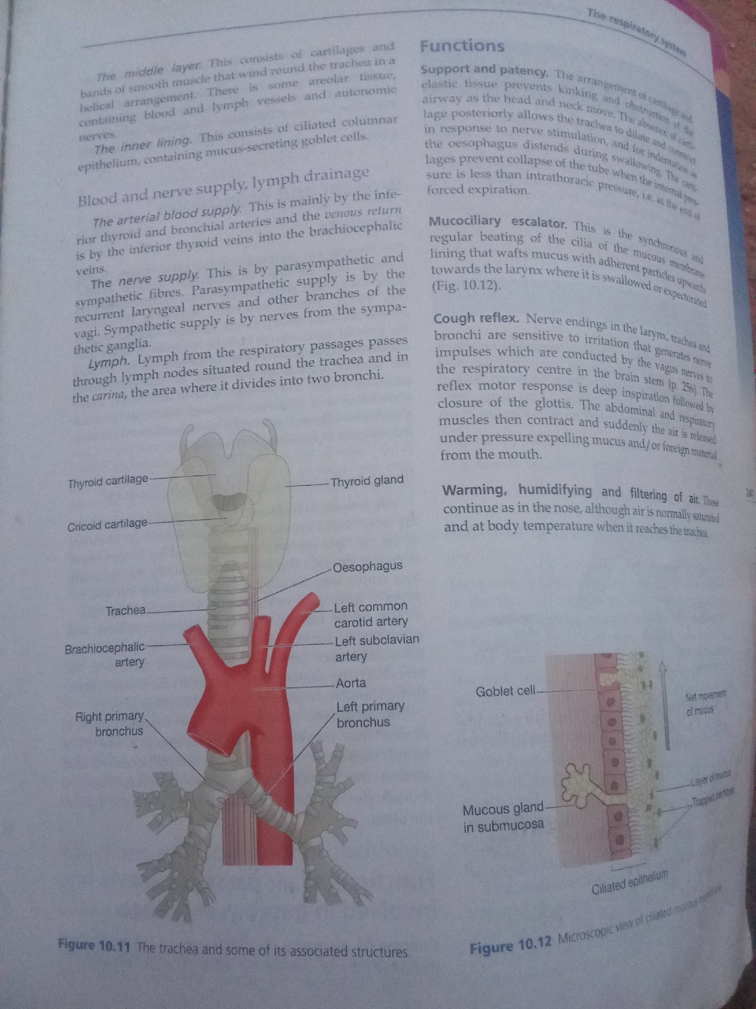
Stake attention in this memory
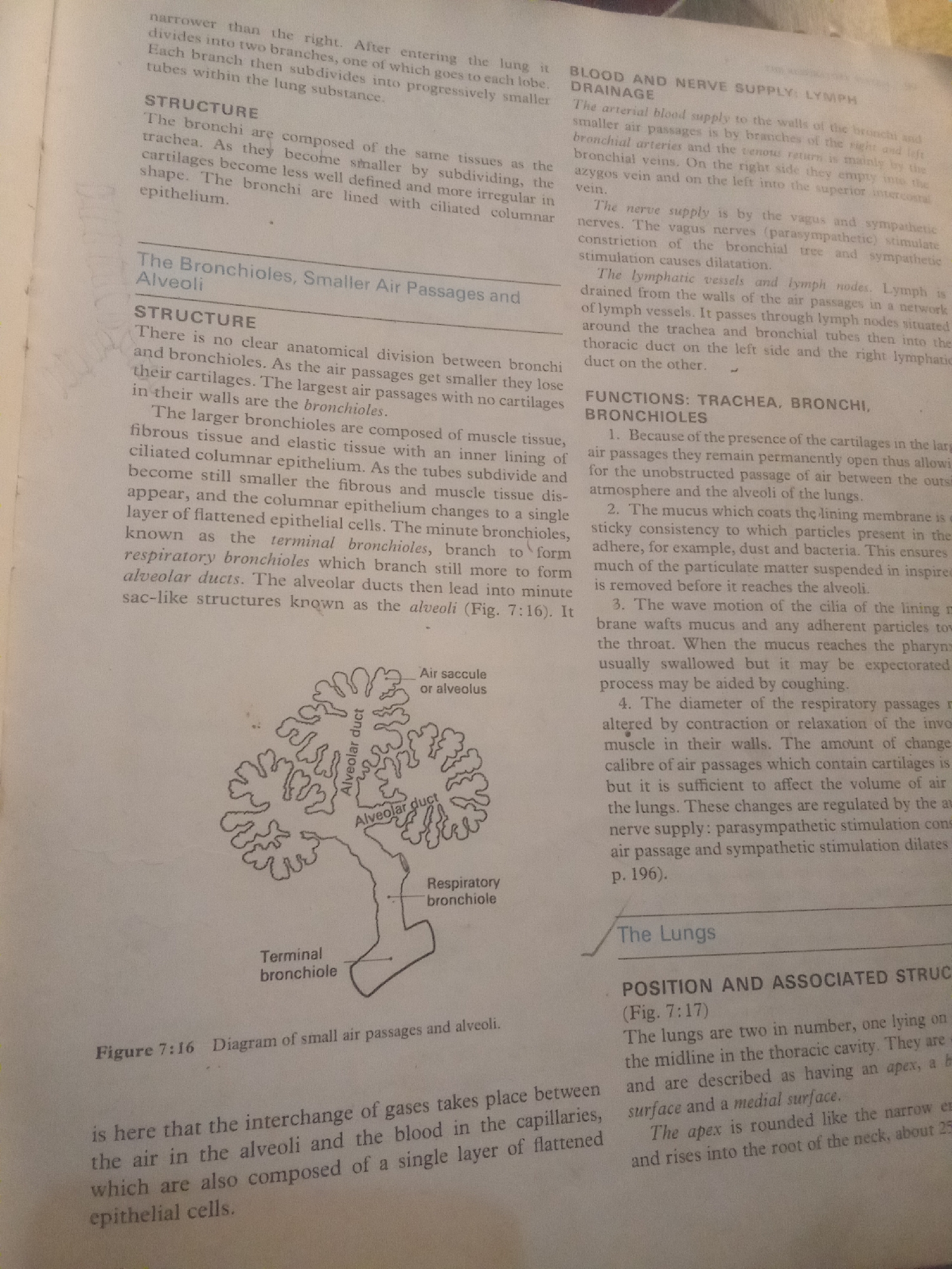
The image is a scanned page from a textbook, likely about human anatomy or biology. It features detailed diagrams and text explaining the structure and function of the respiratory system, specifically the bronchioles, alveoli, and lungs. The main visual elements are: - **Diagrams:** A detailed illustration of terminal bronchioles, alveolar ducts, and alveoli is prominently displayed, showing the branching structures of the lungs. - **Text:** Extensive textual information is present, divided into sections like "STRUCTURE," "BLOOD AND NERVE SUPPLY: LYMPH DRAINAGE," and "FUNCTIONS." The text describes the histological composition of airways, their blood and nerve supply, and their roles in respiration. Some text is in bold headings. Specific text visible includes: - Section titles: "The Bronchioles, Smaller Air Passages and Alveoli," "STRUCTURE," "BLOOD AND NERVE SUPPLY: LYMPH DRAINAGE," "FUNCTIONS: TRACHEA, BRONCHI, BRONCHIOLES," and "The Lungs." - Labels on the diagram: "Terminal bronchiole," "Alveolar duct," "Air saccule or alveolus," and "Respiratory bronchiole." - Figure caption: "Figure 7:16 Diagram of small air passages and alveoli." - Other descriptive text: "narrower than the right. After entering the lung it divides into two branches...", "The arterial blood supply...", "The nerve supply is by the vagus and sympathetic nerves...", "The lymphatic vessels and lymph nodes...", and "The lungs are two in number...". There are no people, animals, or outdoor scenes. The setting is clearly an academic or educational context, with the textbook page being the sole subject. The time of day, weather, and emotional cues are not applicable. The image is of a printed document, likely created in an indoor, well-lit environment for the purpose of scanning.
No transactions found