Stake attention in this memory
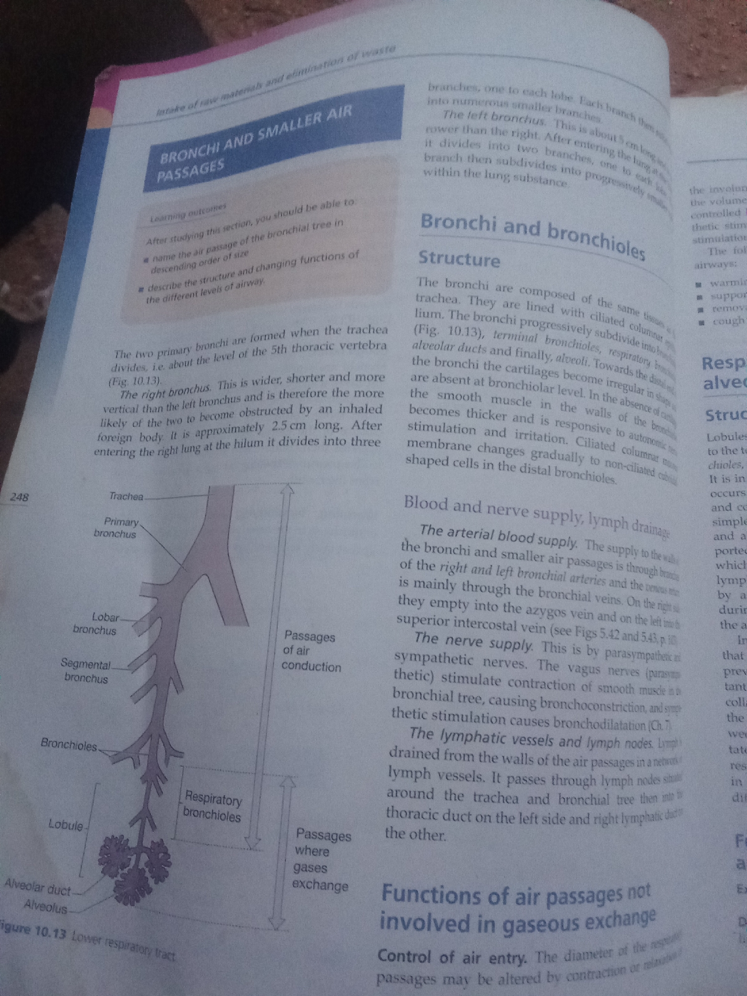
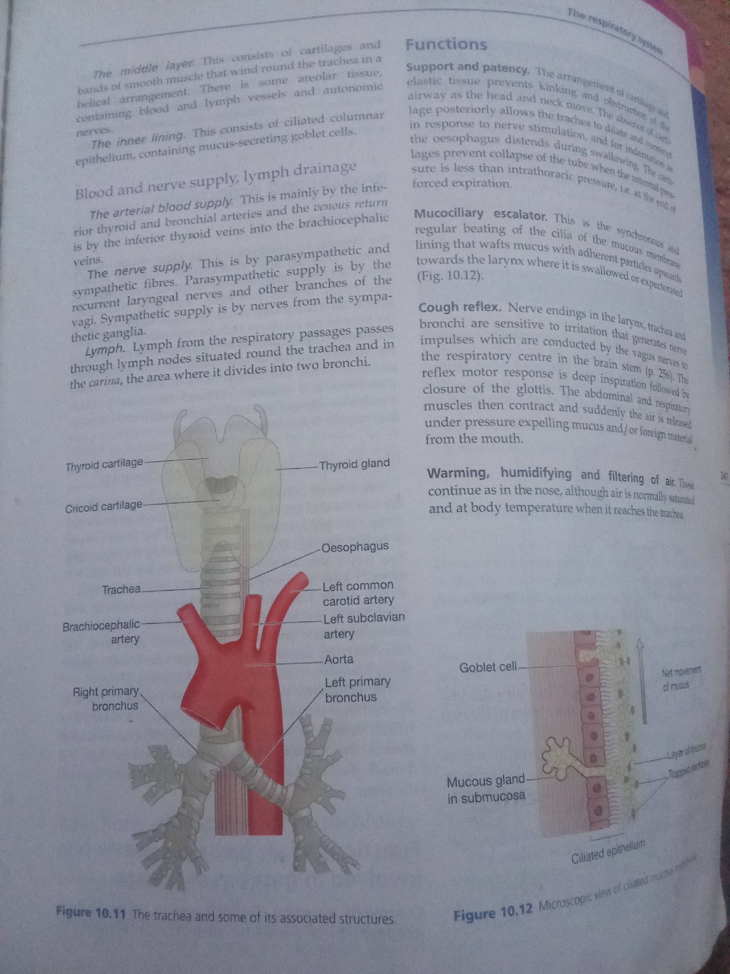
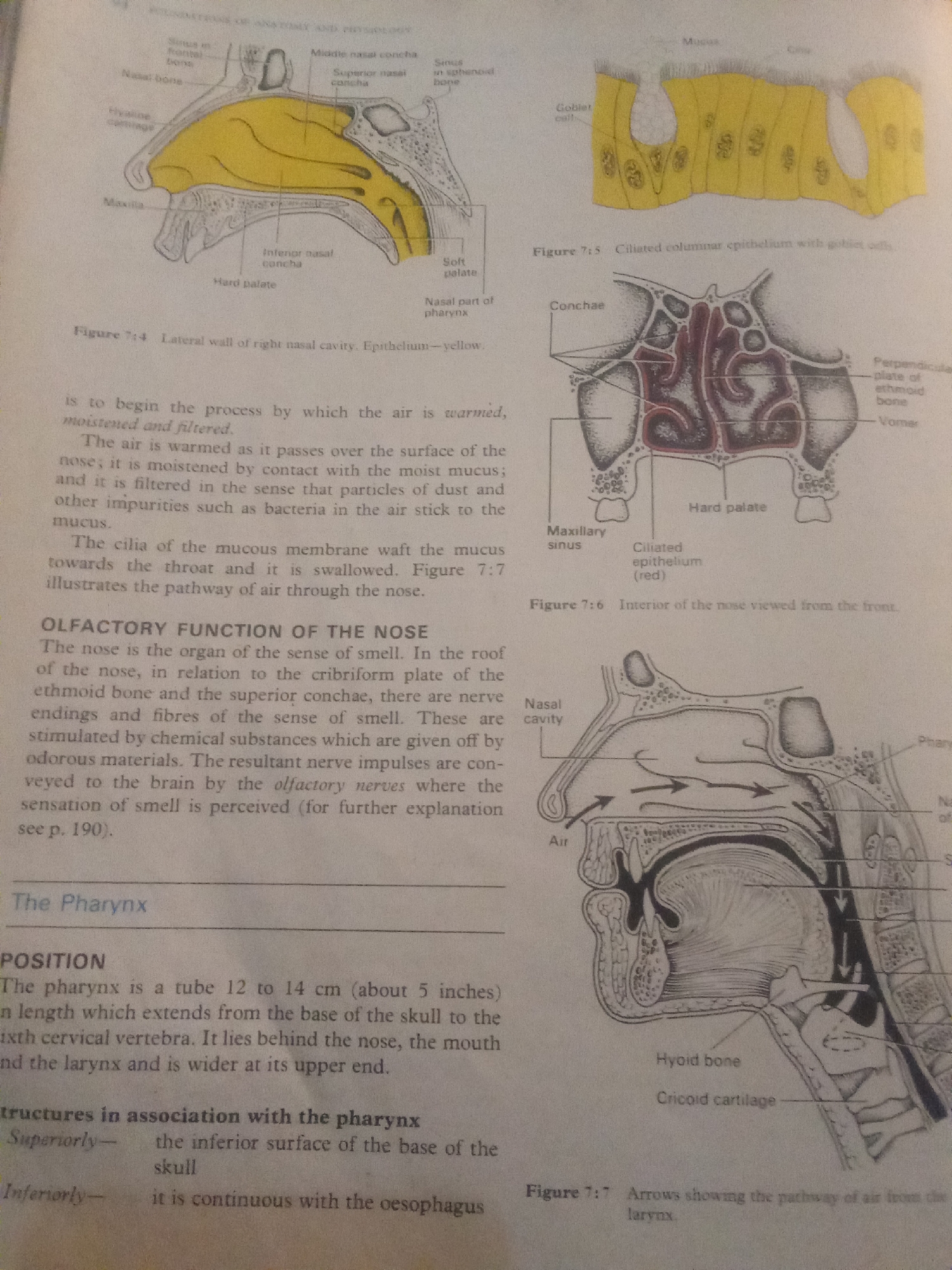
This media file contains diagrams and text from a biology textbook, likely pertaining to the human respiratory and olfactory systems. **Subjects and Objects:** The primary subjects are anatomical illustrations of the nasal cavity, pharynx, and related structures. These include depictions of bones (maxilla, ethmoid, vomer), cartilage (hyaline, cricoid), soft palate, conchae, sinuses, and epithelial cells (ciliated columnar and goblet cells). Arrows indicate the pathway of air. **Activity or Event:** The diagrams illustrate biological processes and anatomical relationships. Specifically, they show the internal structure of the nasal cavity, the arrangement of conchae, the epithelial lining of the respiratory tract, and the passage of air through the upper respiratory system. The text describes the functions of the nose, including warming, moistening, and filtering air, as well as the olfactory sense. It also details the position and structural associations of the pharynx. **Notable Details:** The illustrations are black and white line drawings with some areas highlighted in yellow and red. The yellow highlights likely represent mucus or a specific tissue type in the nasal cavity, while the red highlights a specific epithelial type. The text is printed, suggesting it's from a book or publication. There are no discernible cues about time of day, weather, emotions, or specific location within Jalingo, Nigeria, as this is a generic biological illustration. **Visible Text:** Several labels and captions are visible, including: - "TOY AND PHYSIOLOUT" (likely part of a heading) - Anatomical labels such as "Middle nasal concha," "Superior nasal concha," "Inferior nasal concha," "Hard palate," "Soft palate," "Nasal bone," "Maxilla," "Hyaline cartilage," "Sinus in sphenoid bone," "Goblet cell," "Mucus," "Conchae," "Maxillary sinus," "Ciliated epithelium (red)," "Perpendicular plate of ethmoid bone," "Vomer," "Nasal cavity," "Pharynx," "Hyoid bone," "Cricoid cartilage," and "Air." - Figure captions: "Figure 7:4 Lateral wall of right nasal cavity. Epithelium-yellow.", "Figure 7:5 Ciliated columnar epithelium with goblet cells.", "Figure 7:6 Interior of the nose viewed from the front", "Figure 7:7 Arrows showing the pathway of air from the larynx." - Text describing the function of the nose and the pharynx, including its "POSITION" and "Structures in association with the pharynx." - A reference "see p. 190."
Loading AttnAds…
No transactions found