
Sign in
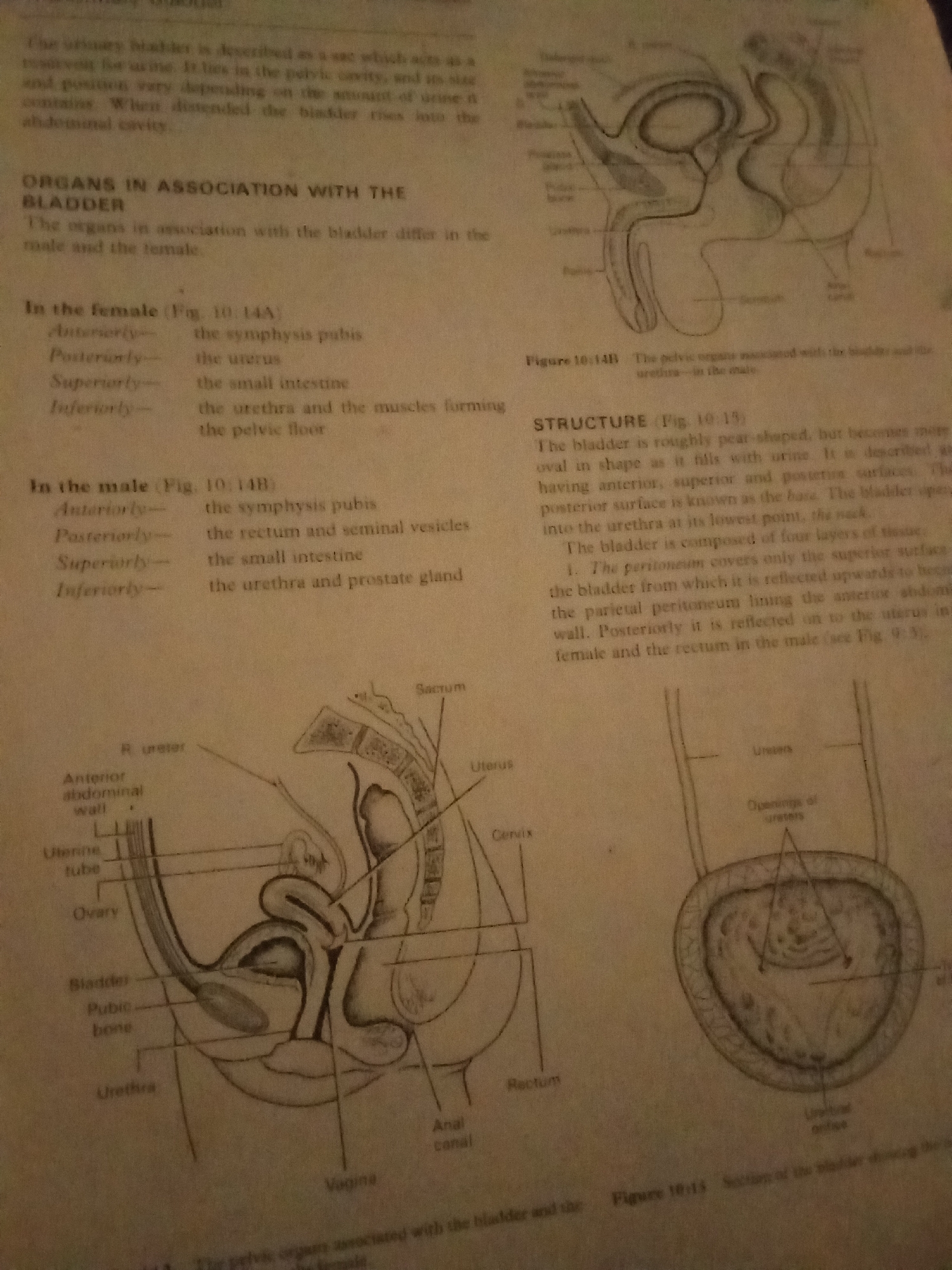
The image displays anatomical illustrations related to the male and female reproductive and urinary systems, along with descriptive text.

The upper left section contains text describing the urinary bladder. The "ORGANS IN ASSOCIATION WITH THE BLADDER" section details associated organs in both females and males. The "In the female" section lists the symphysis pubis, uterus, small intestine, and the urethra and pelvic floor muscles. The "In the male" section lists the symphysis pubis, rectum and seminal vesicles, small intestine, and the urethra and prostate gland.

The "STRUCTURE" section describes the bladder's pear shape, its surfaces, and its four layers of tissue, mentioning the peritoneum, parietal peritoneum, uterus, and rectum.

The image includes two main anatomical diagrams:

1.  **Female Pelvic Anatomy:** A sagittal view of the female pelvis, showing the bladder, pubic bone, urethra, vagina, cervix, uterus, uterine tube, ovary, anterior abdominal wall, and sacrum.
2.  **Male Pelvic Anatomy (Diagrammatic):** A diagram showing the ureters entering the bladder and a cross-section of the bladder, indicating openings of the ureters and the urethral orifice. Another diagram, labeled "Figure 10.14B," shows a general outline of the male pelvic organs.

There is no information within the image that relates to Jalingo, Nigeria.
FM-KpyEx1

Jun 30, 2026, 3:36 PM

Jalingo, Nigeria

Stake attention in this memory

informative
educational
anatomical
scientific
technical

The image displays anatomical illustrations related to the male and female reproductive and urinary systems, along with descriptive text. The upper left section contains text describing the urinary bladder. The "ORGANS IN ASSOCIATION WITH THE BLADDER" section details associated organs in both females and males. The "In the female" section lists the symphysis pubis, uterus, small intestine, and the urethra and pelvic floor muscles. The "In the male" section lists the symphysis pubis, rectum and seminal vesicles, small intestine, and the urethra and prostate gland. The "STRUCTURE" section describes the bladder's pear shape, its surfaces, and its four layers of tissue, mentioning the peritoneum, parietal peritoneum, uterus, and rectum. The image includes two main anatomical diagrams: 1. **Female Pelvic Anatomy:** A sagittal view of the female pelvis, showing the bladder, pubic bone, urethra, vagina, cervix, uterus, uterine tube, ovary, anterior abdominal wall, and sacrum. 2. **Male Pelvic Anatomy (Diagrammatic):** A diagram showing the ureters entering the bladder and a cross-section of the bladder, indicating openings of the ureters and the urethral orifice. Another diagram, labeled "Figure 10.14B," shows a general outline of the male pelvic organs. There is no information within the image that relates to Jalingo, Nigeria.

Loading AttnAds…

transactions
revenues
stakers
Earliest
Latest
Highest stake

No transactions found

More from this user

More from Jalingo

FM-KpyEx1

Jun 30, 2026, 3:36 PM

Jalingo, Nigeria

Licensed under Creative Commons Attribution 4.0 International License. You are free to share and adapt this content with proper attribution.