
Sign in
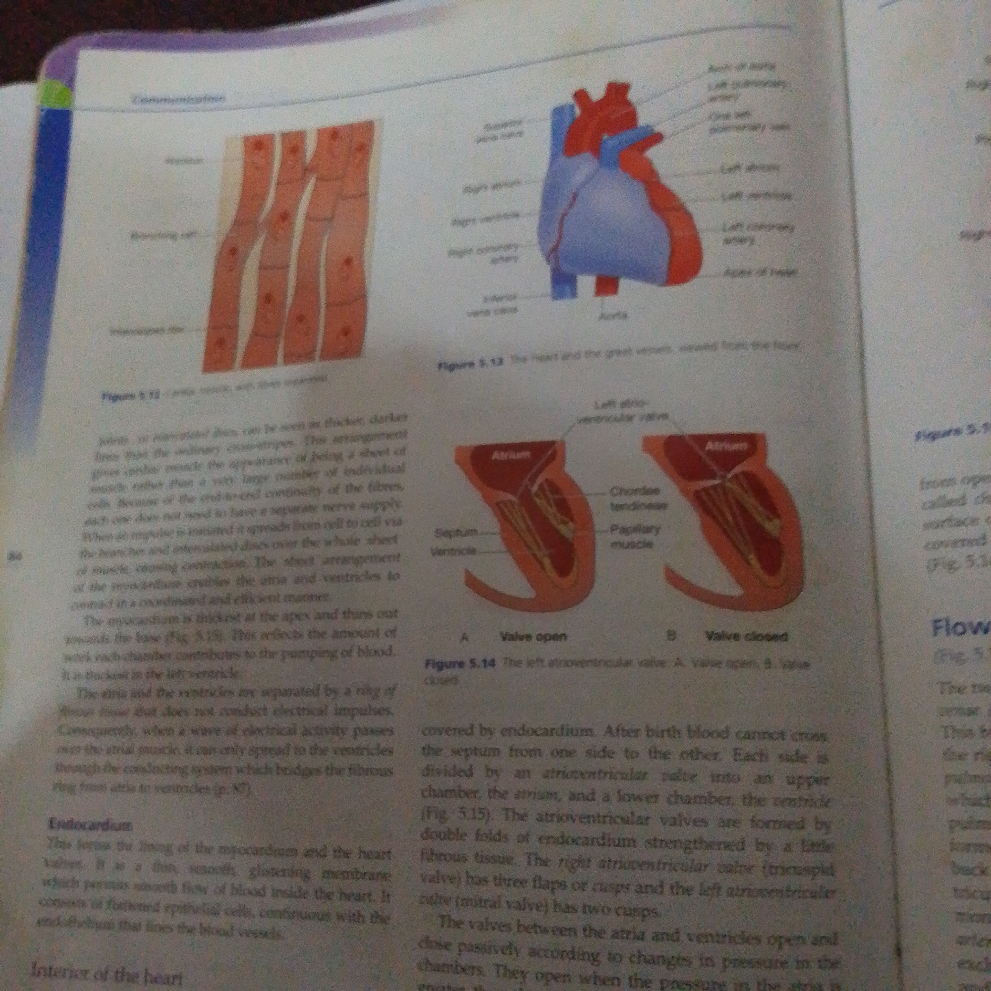
This media file is a photograph of an open textbook page. The page displays diagrams and text related to the anatomy of the human heart and circulatory system.

The subjects are scientific illustrations and written explanations. On the left, there is a diagram of cardiac muscle fibers, labeled with terms like "Atrium" and "Intercalated disc". To the right of this, a larger illustration depicts the human heart, with various parts labeled in red and blue, including the "Right atrium", "Right ventricle", "Left atrium", and "Aorta". Below the heart diagram, a diagram of a heart valve is shown in two states: "A. Valve open" and "B. Valve closed", with labels such as "Septum", "Ventricle", "Chordae tendineae", and "Papillary muscle".

The text discusses the structure and function of cardiac muscle, the heart chambers, and the atrioventricular valves. It explains how electrical impulses spread through the heart and the role of the endocardium. The page also includes captions for the figures, such as "Figure 5.12 Cardiac muscle, with their striations" and "Figure 5.14 The left atrioventricular valve".

There are no people or animals visible in the image. The setting is a book, suggesting an educational context. The overall tone is informative and academic. The lighting appears to be artificial, consistent with indoor study. There are no clear indications of time of day or weather. The visible text is in English and is related to biology and human anatomy.
FM-66btC3

Jun 30, 2026, 1:02 PM

Jalingo, Nigeria

Stake attention in this memory

curious
studious
focused
analytical
inspired

This media file is a photograph of an open textbook page. The page displays diagrams and text related to the anatomy of the human heart and circulatory system. The subjects are scientific illustrations and written explanations. On the left, there is a diagram of cardiac muscle fibers, labeled with terms like "Atrium" and "Intercalated disc". To the right of this, a larger illustration depicts the human heart, with various parts labeled in red and blue, including the "Right atrium", "Right ventricle", "Left atrium", and "Aorta". Below the heart diagram, a diagram of a heart valve is shown in two states: "A. Valve open" and "B. Valve closed", with labels such as "Septum", "Ventricle", "Chordae tendineae", and "Papillary muscle". The text discusses the structure and function of cardiac muscle, the heart chambers, and the atrioventricular valves. It explains how electrical impulses spread through the heart and the role of the endocardium. The page also includes captions for the figures, such as "Figure 5.12 Cardiac muscle, with their striations" and "Figure 5.14 The left atrioventricular valve". There are no people or animals visible in the image. The setting is a book, suggesting an educational context. The overall tone is informative and academic. The lighting appears to be artificial, consistent with indoor study. There are no clear indications of time of day or weather. The visible text is in English and is related to biology and human anatomy.

Loading AttnAds…

transactions
revenues
stakers
Earliest
Latest
Highest stake

No transactions found

More from this user

More from Jalingo

FM-66btC3

Jun 30, 2026, 1:02 PM

Jalingo, Nigeria

Licensed under Creative Commons Attribution 4.0 International License. You are free to share and adapt this content with proper attribution.