Stake attention in this memory
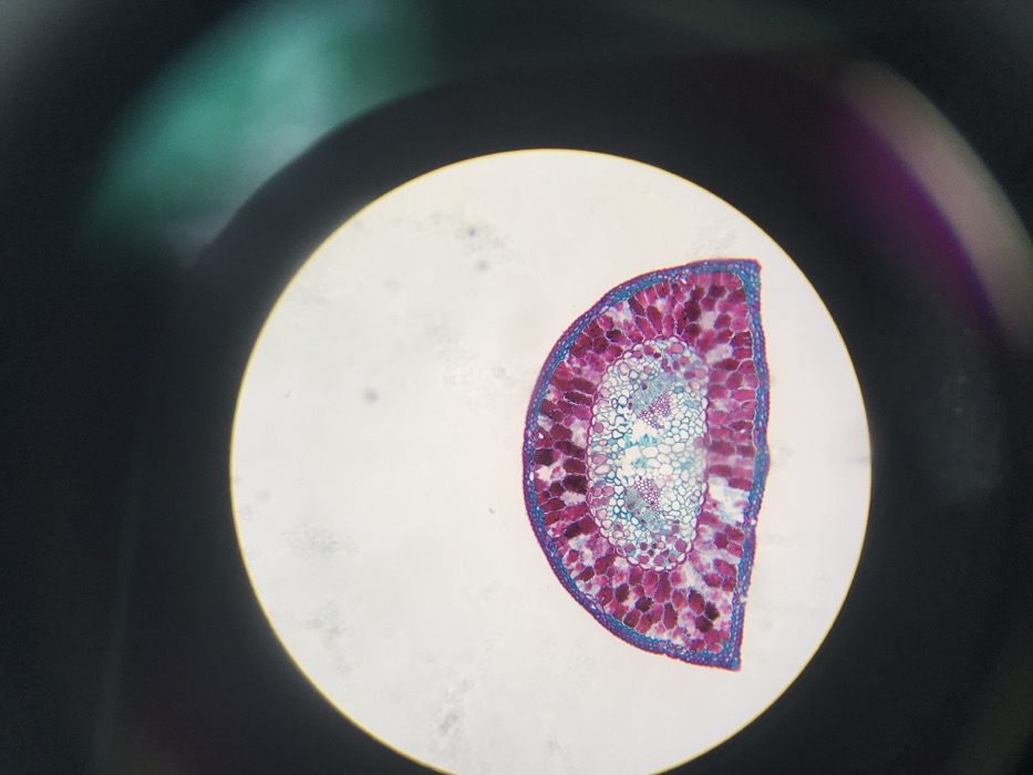
The image displays a microscopic view of a stained biological cross-section, presented within a circular, brightly illuminated field. This circular field occupies the central and lower-middle portions of the frame, surrounded by a dark, out-of-focus periphery that exhibits subtle lens aberrations, including faint greenish-blue and reddish-purple flares. The primary subject within the illuminated circle is a semi-circular cross-section of plant tissue, positioned towards the bottom of the field. The tissue is stained, revealing distinct cellular structures and differentiation. The outermost layers of the tissue exhibit a deep purple to dark pink coloration, consistent with epidermal and cortical cells. Interior to these, a more compact layer of cells is stained a vibrant magenta. The central vascular region is characterized by an oval-shaped cluster of cells, stained a lighter, bluish-green, indicating xylem and phloem bundles. No human figures or direct actions are depicted. Several small, dark specks, likely dust or debris, are visible within the bright circular field. The specific geographic location in Midrand, South Africa, cannot be determined from the visual content of this microscopic image.
No transactions found