Stake attention in this memory
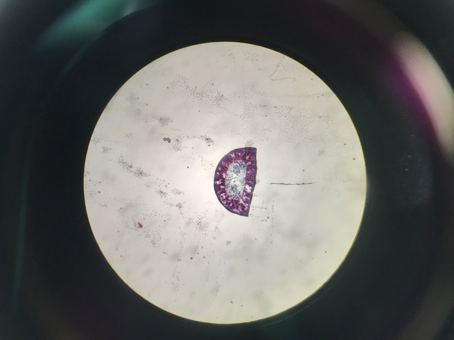
This image captures a microscopic cross-section of a stained plant tissue, specifically a conifer needle, as viewed through a microscope eyepiece in a laboratory setting in Midrand, South Africa. The biological sample occupies approximately one-third of the central circular field of view, exhibiting a distinct semi-circular morphology. The tissue displays various cellular structures stained predominantly in magenta and light blue-green, with a darker magenta outer layer enclosing an inner core featuring lighter blue-green vascular bundles. The white-grey background within the circular observation field contains numerous fine dark specks and larger irregular dark particles, consistent with dust or debris on the slide or optical lenses. The area immediately outside the circular field is dark, with incidental reflections of green and magenta light visible on the periphery. No individuals, direct actions, or interactions are visible in the frame. The scene represents a static record of a scientific observation.
No transactions found