Sign in
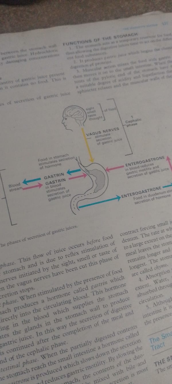
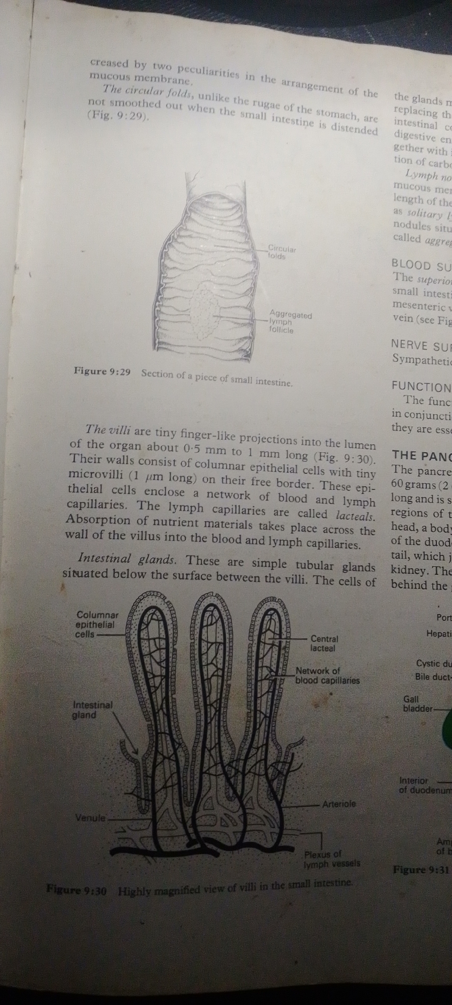
This media file contains a scanned page from a biology textbook, detailing the anatomy of the small intestine. The page features text and two diagrams.

The main subject of the page is the structure and function of the small intestine. The text describes the circular folds, villi, and intestinal glands, explaining their roles in digestion and absorption. It also mentions blood and lymph supply, as well as nerve supply.

The first diagram, labeled Figure 9:29, is a cross-section of a piece of the small intestine, illustrating circular folds and an aggregated lymph follicle. The second diagram, Figure 9:30, is a highly magnified view of villi within the small intestine, showing columnar epithelial cells, blood capillaries, and lacteals. A third, partially visible diagram on the right side of the page (Figure 9:31) appears to depict internal organs, with labels like "Interior of duodenum" and "Gall bladder."

There are no people, animals, or natural scenes depicted. The setting is academic, indicated by the textbook format and scientific illustrations. The activity depicted is purely illustrative, explaining biological concepts through diagrams and text. There are no visible cues regarding time of day, weather, or emotions.

The visible text includes: "creased by two peculiarities in the arrangement of the mucous membrane," "The circular folds, unlike the rugae of the stomach, are not smoothed out when the small intestine is distended (Fig. 9:29)," "Figure 9:29 Section of a piece of small intestine," "The villi are tiny finger-like projections into the lumen of the organ about 0.5 mm to 1 mm long (Fig. 9:30)," "Intestinal glands. These are simple tubular glands situated below the surface between the villi," "Columnar epithelial cells," "Intestinal gland," "Venule," "Central lacteal," "Network of blood capillaries," "Arteriole," "Plexus of lymph vessels," "Figure 9:30 Highly magnified view of villi in the small intestine," and text related to the pancreas and other organs on the right side.
FM-2L6Lq2

Jun 30, 2026, 4:49 PM

Jauro Yinu, Nigeria

Moment

Stake attention in this memory

Curiosity
Educational
Analytical
Clinical
Focused

This media file contains a scanned page from a biology textbook, detailing the anatomy of the small intestine. The page features text and two diagrams. The main subject of the page is the structure and function of the small intestine. The text describes the circular folds, villi, and intestinal glands, explaining their roles in digestion and absorption. It also mentions blood and lymph supply, as well as nerve supply. The first diagram, labeled Figure 9:29, is a cross-section of a piece of the small intestine, illustrating circular folds and an aggregated lymph follicle. The second diagram, Figure 9:30, is a highly magnified view of villi within the small intestine, showing columnar epithelial cells, blood capillaries, and lacteals. A third, partially visible diagram on the right side of the page (Figure 9:31) appears to depict internal organs, with labels like "Interior of duodenum" and "Gall bladder." There are no people, animals, or natural scenes depicted. The setting is academic, indicated by the textbook format and scientific illustrations. The activity depicted is purely illustrative, explaining biological concepts through diagrams and text. There are no visible cues regarding time of day, weather, or emotions. The visible text includes: "creased by two peculiarities in the arrangement of the mucous membrane," "The circular folds, unlike the rugae of the stomach, are not smoothed out when the small intestine is distended (Fig. 9:29)," "Figure 9:29 Section of a piece of small intestine," "The villi are tiny finger-like projections into the lumen of the organ about 0.5 mm to 1 mm long (Fig. 9:30)," "Intestinal glands. These are simple tubular glands situated below the surface between the villi," "Columnar epithelial cells," "Intestinal gland," "Venule," "Central lacteal," "Network of blood capillaries," "Arteriole," "Plexus of lymph vessels," "Figure 9:30 Highly magnified view of villi in the small intestine," and text related to the pancreas and other organs on the right side.

Symbol

336EA

Volume

4,792

Creator

+$0.01

Revenue

+$0.01

TVL

$0.54

1D
1W
1M
All

Loading AttnAds…

transactions
revenues
stakers
Earliest
Latest
Highest stake

1

FM-FSRgn1

Staked 16d ago

$0.54 USD

+$0.00 USD

+0.4%

2

Rev Bot 🤖💰

Injected revenue 16d ago

Revenue bonus for the last stake.

+$0.01 USD

More from this user

More from Jauro Yinu

FM-2L6Lq2

Jun 30, 2026, 4:49 PM

Jauro Yinu, Nigeria

Licensed under Creative Commons Attribution 4.0 International License. You are free to share and adapt this content with proper attribution.