
Stake attention in this memory
anatomy
liver
medical illustration
biology
histology
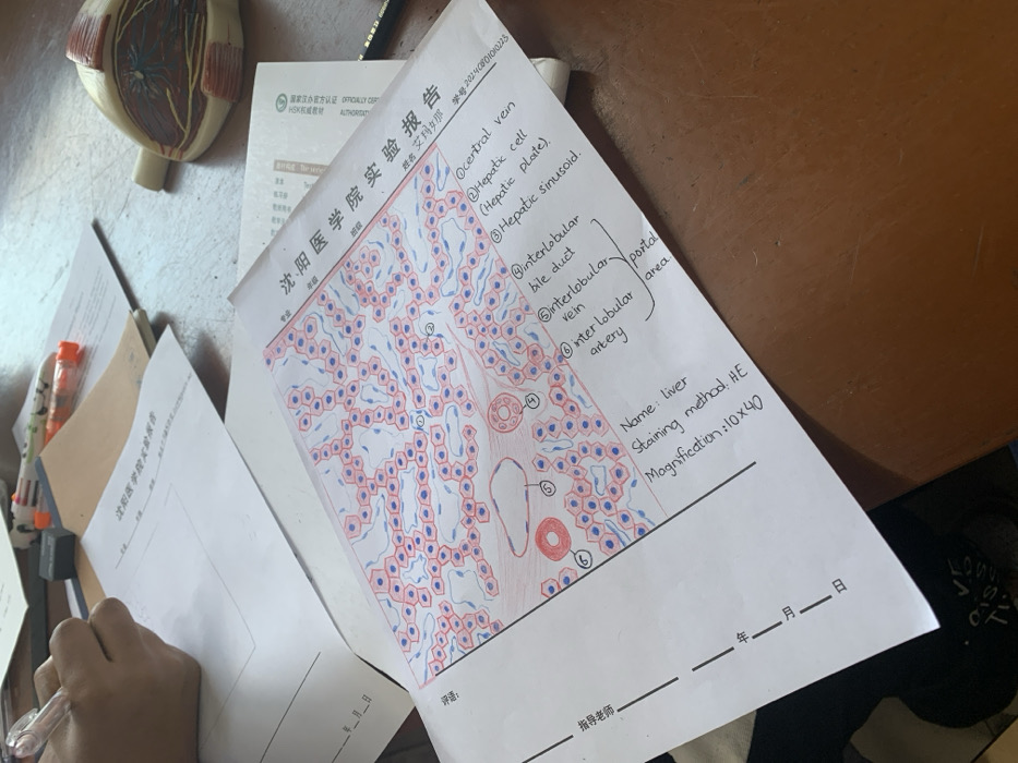
The image shows a drawing of a liver's microscopic structure, labeled with corresponding terms. A "central vein," "hepatic cell (hepatic plate), hepatic sinusoid," and "portal area" are indicated. Within the portal area, labels include "interlobular bile duct," "interlobular vein," and "interlobular artery." At the bottom right of the image, it's noted that the sample is a "Liver" sample, "staining method: HE," and "Magnification: 10x40." A student's name, student ID, and grade are listed at the top of the image. The form is a lab report from Shenyang Medical College.
Loading AttnAds…
transactions
revenues
stakers
Earliest
Latest
Highest stake
No transactions found