
Stake attention in this memory
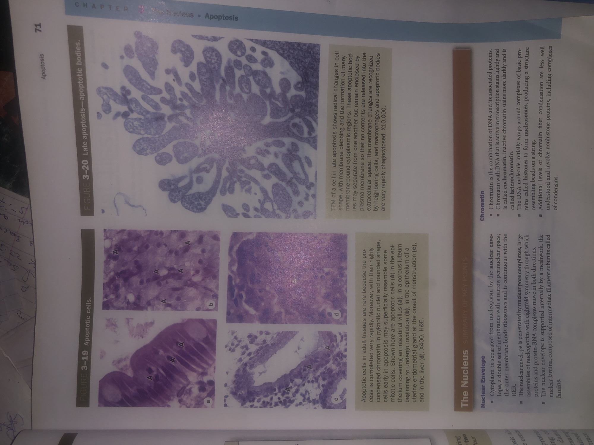
The image is a scan of a textbook page discussing apoptosis. It features two main figures, labeled Figure 3-19 and Figure 3-20, which are microscopic images related to apoptotic cells and apoptotic bodies. Figure 3-19 shows four smaller photomicrographs labeled a, b, c, and d. These images depict apoptotic cells (labeled "A") in various tissues, including the epithelium of an intestinal villus, a corpus luteum, an epithelial gland, and the liver. The accompanying text provides a detailed explanation of these images and the characteristics of apoptotic cells. Figure 3-20 displays a Transmission Electron Microscope (TEM) image of a cell in late apoptosis, highlighting membrane blebbing and the formation of apoptotic bodies. The caption explains the ultrastructural changes observed and the rapid phagocytosis of these bodies. Below the figures, there are sections titled "The Nucleus SUMMARY OF KEY POINTS" and "Chromatin," which further elaborate on nuclear structure and chromatin organization. The text also includes page numbers and chapter information, indicating this is likely from a biology or histology textbook. There is no information within the image or its content that relates to the city of Wukari, Nigeria, or any specific location. The description is purely scientific and educational.
Symbol
39F94
Volume
11,463
Creator
+$0.03
Revenue
+$0.06
TVL
$2.83
Loading AttnAds…

2

Rev Bot 🤖💰
Injected revenue 27d ago
“Revenue bonus for the last stake.”
+$0.07 USD