Stake attention in this memory
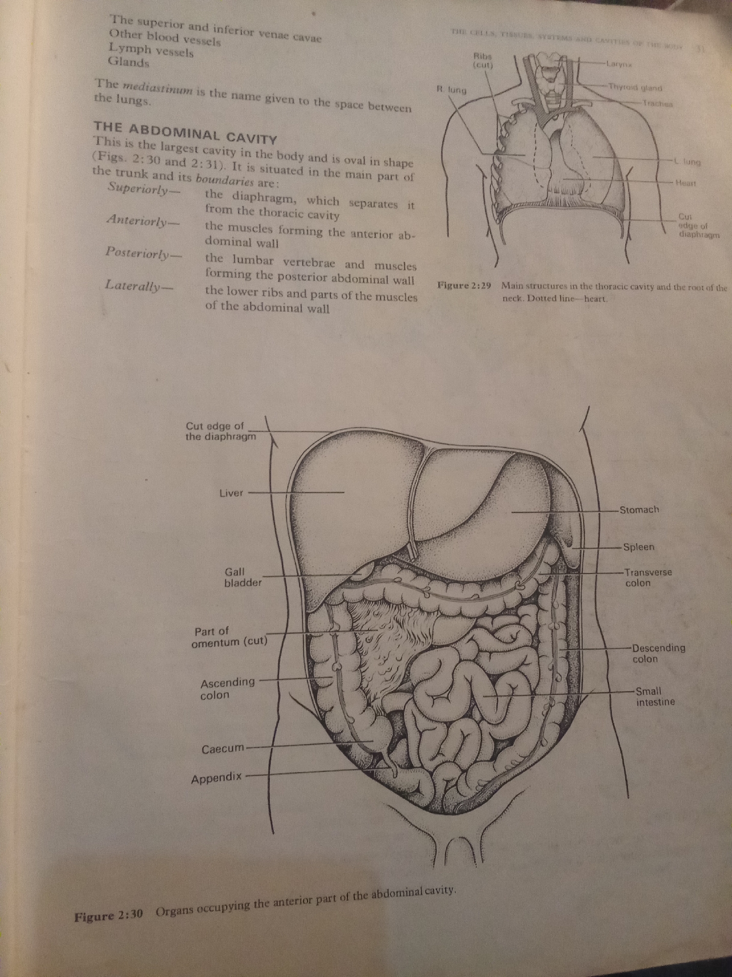
This media file is a page from an anatomy textbook, featuring black and white illustrations of the human torso. The primary subjects are anatomical diagrams of the thoracic and abdominal cavities. The top of the page shows Figure 2:29, illustrating the main structures of the thoracic cavity, including the lungs, heart, trachea, larynx, and thyroid gland. Text labels these various organs and anatomical features. Below this is text describing the mediastinum and the abdominal cavity, defining its boundaries: superiorly by the diaphragm, anteriorly by abdominal wall muscles, posteriorly by lumbar vertebrae and muscles, and laterally by the lower ribs and abdominal muscles. The lower portion of the page displays Figure 2:30, a detailed diagram of the abdominal cavity, showing organs such as the liver, gall bladder, stomach, spleen, transverse colon, descending colon, small intestine, caecum, and appendix. Labels point to these organs and structures, including the cut edge of the diaphragm. The caption for Figure 2:30 states it depicts "Organs occupying the anterior part of the abdominal cavity." The visible text provides anatomical and descriptive information related to the illustrations. There are no people, discernible emotions, or specific time of day or weather cues as it is a textbook page. The location context provided (Jalingo, Nigeria) is external to the image content itself. The media is purely informational and educational.
No transactions found