Stake attention in this memory
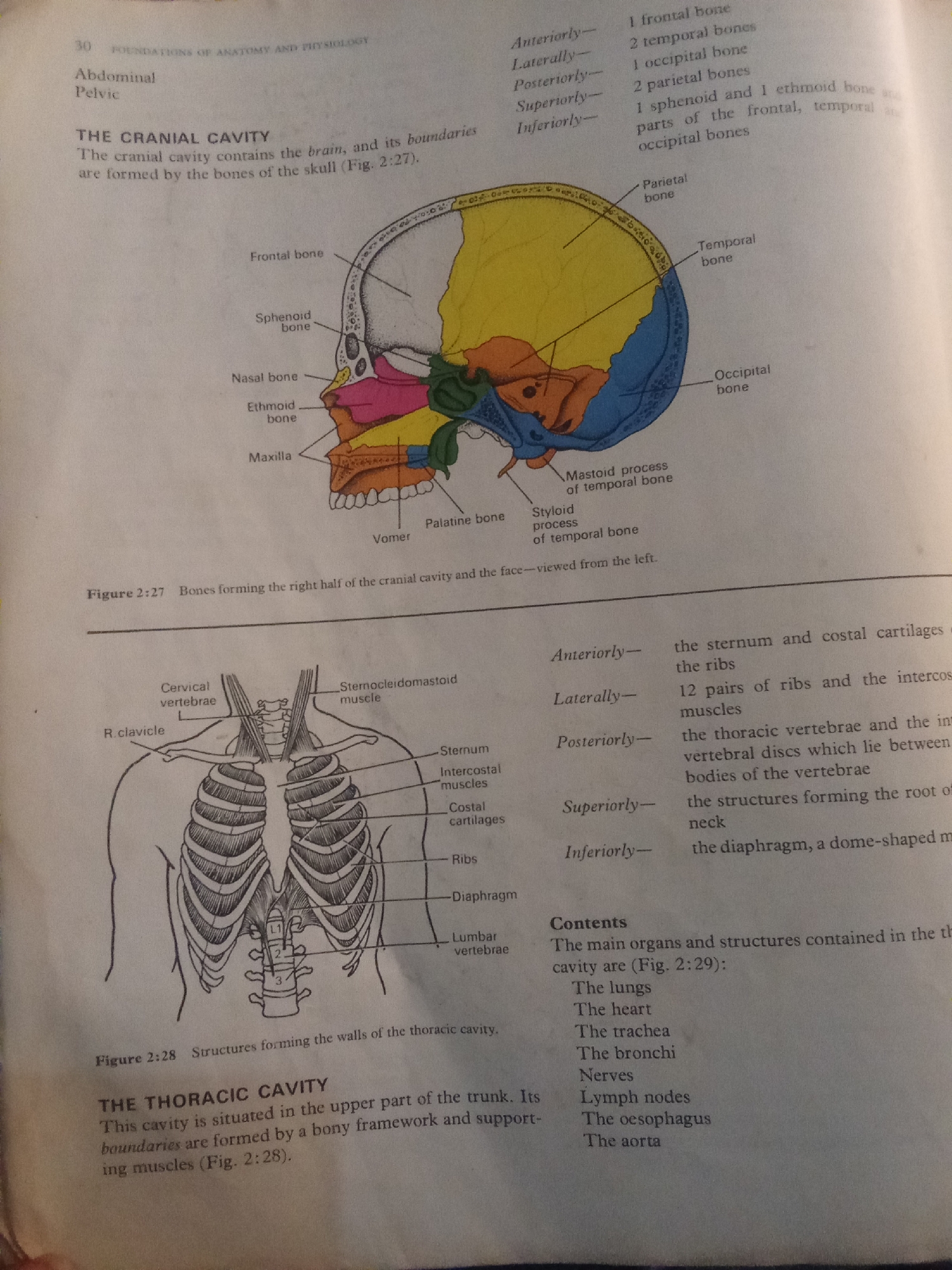
This is an image of a page from an anatomy textbook. The page discusses the cranial and thoracic cavities. The upper half of the image displays a diagram of the human skull, illustrating the bones that form the cranial cavity. Labels point to various bones such as the frontal, sphenoid, nasal, ethmoid, maxilla, parietal, and temporal bones. Text beside the diagram lists the bones that form the cranial cavity anteriorly, laterally, posteriorly, superiorly, and inferiorly. The lower half of the image shows a diagram of the human rib cage and upper spine. Labels indicate structures like the cervical vertebrae, clavicle, sternocleidomastoid muscle, sternum, intercostal muscles, costal cartilages, ribs, diaphragm, and lumbar vertebrae. Below this diagram, a section titled "THE THORACIC CAVITY" describes its location and boundaries, referencing Figure 2:28. A "Contents" list enumerates the main organs and structures found within the thoracic cavity, including the lungs, heart, trachea, bronchi, nerves, lymph nodes, oesophagus, and aorta. The image is likely from a book or educational material used in Jalingo, Nigeria, given the provided location context. There are no people, specific weather conditions, or time of day indicated. The focus is purely on anatomical illustrations and descriptive text.
No transactions found