
Stake attention in this memory
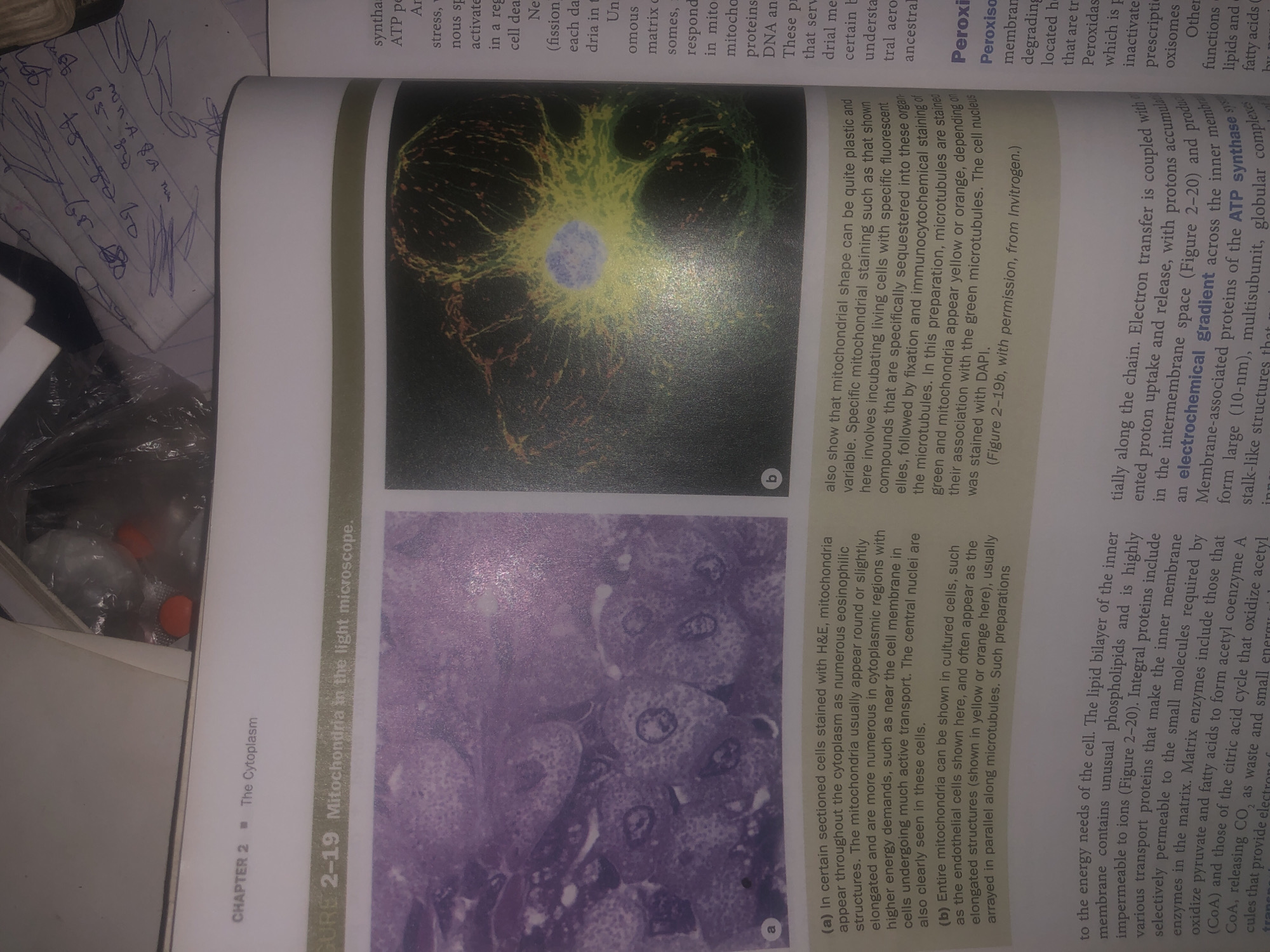
The image displays two micrographs of mitochondria within cells. Micrograph (a) shows mitochondria in sectioned cells stained with H&E, appearing as numerous eosinophilic structures throughout the cytoplasm. They are described as usually round or slightly elongated and more numerous in regions with higher energy demands, such as near the cell membrane. Micrograph (b) presents a cultured cell showing entire mitochondria, appearing as elongated structures in yellow or orange, arranged in parallel along microtubules which are stained green. The cell nucleus is stained blue. The text also discusses mitochondrial shape plasticity and specific fluorescent staining techniques. The image is from a textbook, as indicated by the chapter and figure numbering. The provided crops show parts of the book page, including the chapter title "CHAPTER 2 - The Cytoplasm" and the figure title "FIGURE 2-19 Mitochondria in the light microscope." There is no information in the image that relates to Wukari, Nigeria.
Symbol
43C32
Volume
3,307
Creator
+$0.00
Revenue
+$0.00
TVL
$0.27
Loading AttnAds…