
Stake attention in this memory
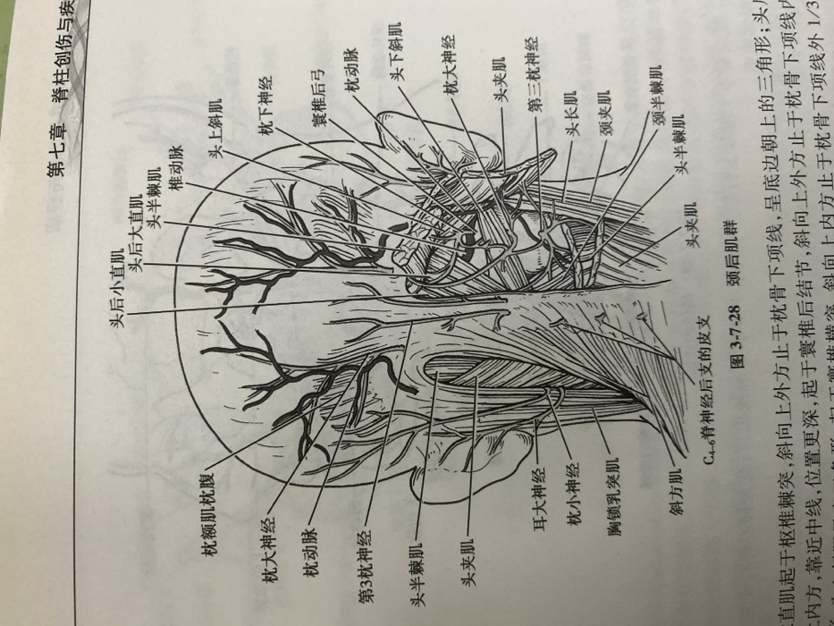
This anatomical illustration depicts the posterior muscles and nerves of the head and neck. Key structures labeled include the occipitofrontalis muscle (occipital belly), greater occipital nerve, occipital artery, third occipital nerve, semispinalis capitis, splenius capitis, greater auricular nerve, lesser occipital nerve, sternocleidomastoid muscle, and trapezius muscle. Also shown are the rectus capitis posterior minor and major, superior oblique, inferior oblique, lesser occipital nerve, greater occipital nerve, splenius capitis, longissimus capitis, spinalis capitis, semispinalis capitis, and splenius capitis. C4-6 posterior branch of cutaneous nerve is identified. A description accompanies a drawing of the posterior cervical muscles.
Loading AttnAds…
No transactions found