Sign in
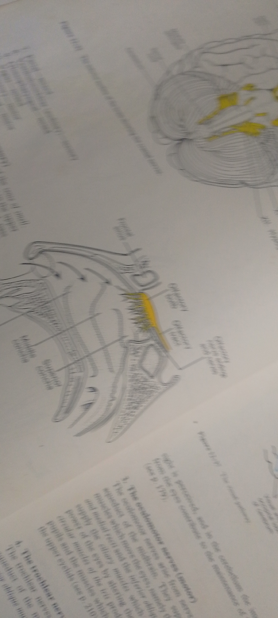
This media file contains a scanned image of a page from a textbook or scientific publication. The primary subjects are anatomical illustrations of the human eye and brain. The illustrations are line drawings with some areas highlighted in yellow. There is also visible text, including captions for the illustrations and descriptive paragraphs.

The illustrations depict the internal structure of the eye, including what appears to be the lens, iris, and surrounding muscles, with labels such as "Pupillary m." and "Ciliary m.". Another illustration shows a cross-section of the brain, with intricate lines representing its structure, and some areas are colored yellow, possibly indicating specific regions or pathways.

The text includes headings like "3. The oculomotor nerves (motor)" and "4. The trochlear nerves", suggesting a focus on the nervous system and its role in eye function. The content discusses the origin and function of these nerves, mentioning their connection to muscles that control eye movement and pupil dilation.

There are no people, specific scene elements, time of day, weather, or discernible emotions visible in this image, as it is a reproduction of printed material. The location context "Jauro Yinu, Nigeria" is provided externally and is not derivable from the image itself. The activity depicted is informational, presenting scientific diagrams and text for educational purposes.
FM-2L6Lq2

Jun 30, 2026, 5:19 PM

Jauro Yinu, Nigeria

Moment

Stake attention in this memory

educational
informative
scientific
detailed
clinical

This media file contains a scanned image of a page from a textbook or scientific publication. The primary subjects are anatomical illustrations of the human eye and brain. The illustrations are line drawings with some areas highlighted in yellow. There is also visible text, including captions for the illustrations and descriptive paragraphs. The illustrations depict the internal structure of the eye, including what appears to be the lens, iris, and surrounding muscles, with labels such as "Pupillary m." and "Ciliary m.". Another illustration shows a cross-section of the brain, with intricate lines representing its structure, and some areas are colored yellow, possibly indicating specific regions or pathways. The text includes headings like "3. The oculomotor nerves (motor)" and "4. The trochlear nerves", suggesting a focus on the nervous system and its role in eye function. The content discusses the origin and function of these nerves, mentioning their connection to muscles that control eye movement and pupil dilation. There are no people, specific scene elements, time of day, weather, or discernible emotions visible in this image, as it is a reproduction of printed material. The location context "Jauro Yinu, Nigeria" is provided externally and is not derivable from the image itself. The activity depicted is informational, presenting scientific diagrams and text for educational purposes.

Symbol

50508

Volume

3,544

Creator

+$0.00

Revenue

+$0.00

TVL

$0.32

1D
1W
1M
All

Loading AttnAds…

transactions
revenues
stakers
Earliest
Latest
Highest stake

1

FM-FSRgn1

Staked 16d ago

$0.32 USD

+$0.00 USD

+0.3%

2

Rev Bot 🤖💰

Injected revenue 16d ago

Revenue bonus for the last stake.

+$0.00 USD

More from this user

More from Jauro Yinu

FM-2L6Lq2

Jun 30, 2026, 5:19 PM

Jauro Yinu, Nigeria

Licensed under Creative Commons Attribution 4.0 International License. You are free to share and adapt this content with proper attribution.