Stake attention in this memory
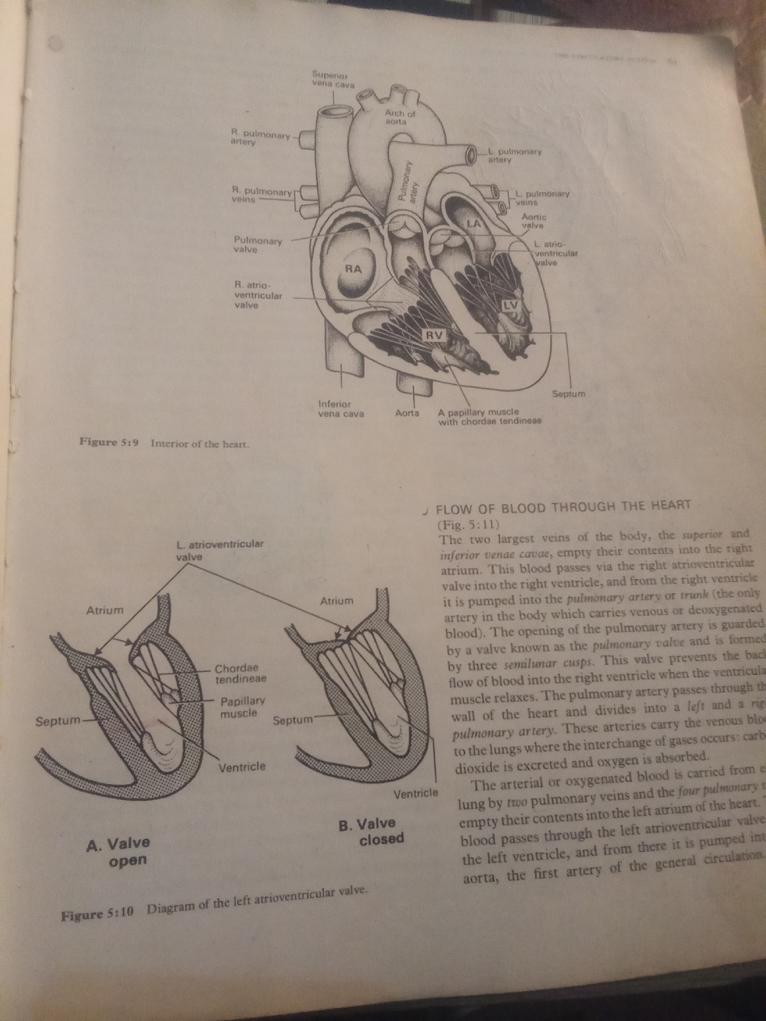
This media file contains an illustration of the human heart with detailed labels for its various parts, including chambers like the right atrium (RA), left atrium (LA), right ventricle (RV), and left ventricle (LV), as well as valves such as the pulmonary valve, aortic valve, and atrioventricular valves. The diagram also shows major blood vessels connected to the heart, like the superior and inferior vena cava, pulmonary artery, and pulmonary veins. Below the main heart diagram, there are two smaller illustrations labeled "A. Valve open" and "B. Valve closed," which specifically depict the mechanism of the left atrioventricular valve, showing the chordae tendineae and papillary muscles. Accompanying the illustrations are text captions and a descriptive paragraph. Figure 5:9 is titled "Interior of the heart," and Figure 5:10 is labeled "Diagram of the left atrioventricular valve." The main text discusses the "Flow of blood through the heart," explaining the path of blood circulation from the veins, through the heart chambers and valves, to the lungs, and back to the heart. It details how deoxygenated blood is pumped to the lungs and how oxygenated blood returns to the left side of the heart before being pumped into the aorta for general circulation. The visible text includes anatomical terms, figure numbers and titles, and a narrative explanation of cardiac function. There are no people, animals, or specific environmental details like weather or time of day depicted; the focus is purely on anatomical and physiological illustration. The context provided for Jalingo, Nigeria, does not align with the content of the image, which is a generic biological diagram.
Loading AttnAds…
No transactions found