
Sign in
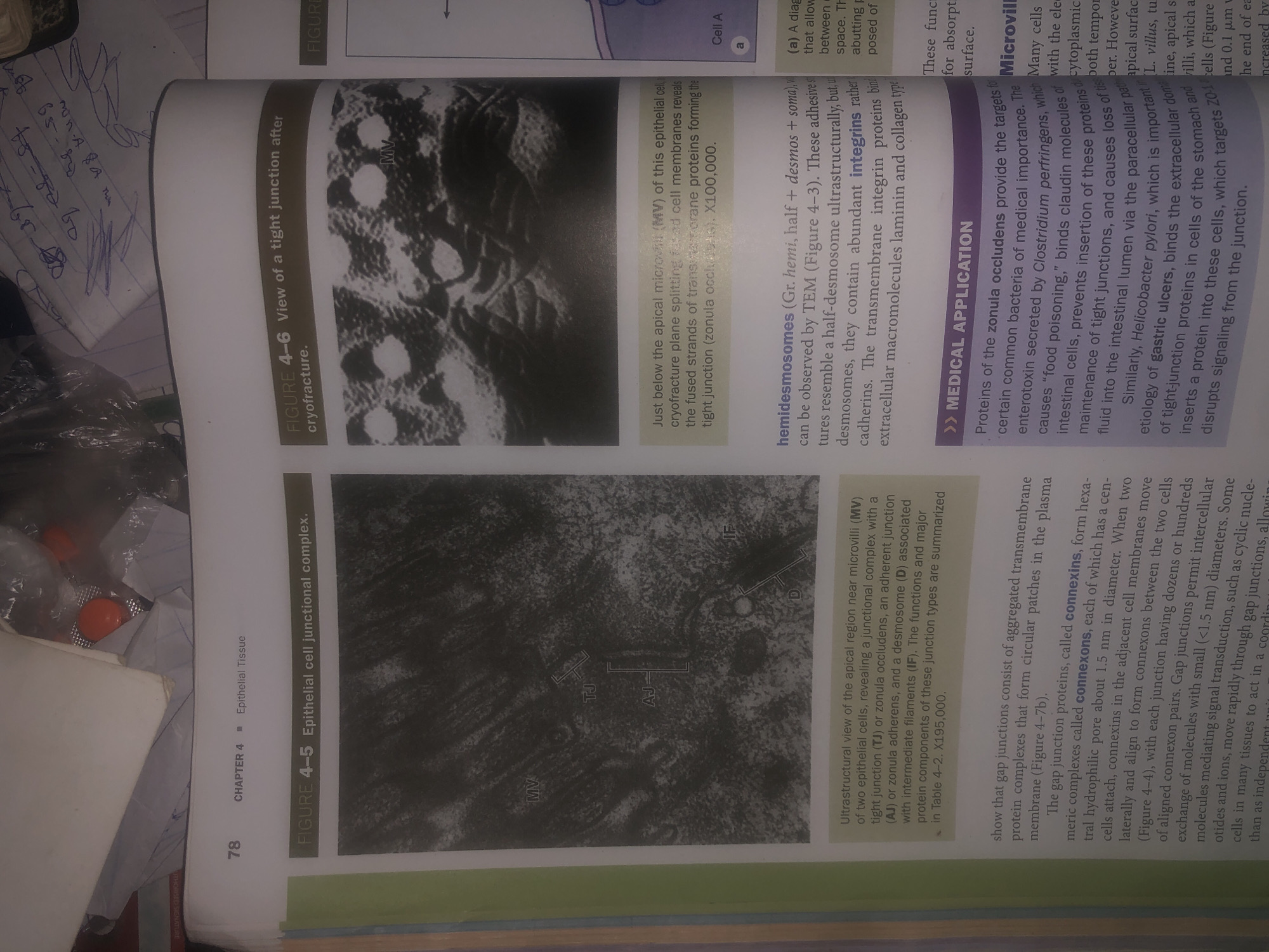
The image is a photograph of an open textbook showing two pages of biological content. The left page displays a micrograph labeled "FIGURE 4-5 Epithelial cell junctional complex." This micrograph shows a close-up view of the junctional complex between two epithelial cells, with labels indicating microvilli (MV), tight junction (TJ), adherens junction (AJ), desmosome (D), and intermediate filaments (IF). Below the micrograph is a detailed description of the ultrastructure of the apical region near microvilli.

The right page features a micrograph labeled "FIGURE 4-6 View of a tight junction after cryofracture." This image appears to be a freeze-fracture electron micrograph showing the strands of transmembrane proteins forming the tight junction. Text below this micrograph explains the view and magnification. Further down the right page, there is a section titled ">> MEDICAL APPLICATION" discussing the role of proteins of the zonula occludens in medical importance, mentioning their interaction with bacteria and the impact on intestinal permeability. Below this, the text discusses hemidesmosomes and the role of integrins.

The overall scene depicted is an academic textbook with scientific illustrations and textual explanations related to epithelial tissue and cell junctions. There are no people, subjects, objects, actions, interactions, or environmental elements that would be relevant to a location in Wukari, Nigeria. The context is purely educational and biological.
Bigstev

Jun 12, 2026, 1:52 AM

Wukari, Nigeria

Short Term

Stake attention in this memory

educational
informative
scientific
complex
detailed

The image is a photograph of an open textbook showing two pages of biological content. The left page displays a micrograph labeled "FIGURE 4-5 Epithelial cell junctional complex." This micrograph shows a close-up view of the junctional complex between two epithelial cells, with labels indicating microvilli (MV), tight junction (TJ), adherens junction (AJ), desmosome (D), and intermediate filaments (IF). Below the micrograph is a detailed description of the ultrastructure of the apical region near microvilli. The right page features a micrograph labeled "FIGURE 4-6 View of a tight junction after cryofracture." This image appears to be a freeze-fracture electron micrograph showing the strands of transmembrane proteins forming the tight junction. Text below this micrograph explains the view and magnification. Further down the right page, there is a section titled ">> MEDICAL APPLICATION" discussing the role of proteins of the zonula occludens in medical importance, mentioning their interaction with bacteria and the impact on intestinal permeability. Below this, the text discusses hemidesmosomes and the role of integrins. The overall scene depicted is an academic textbook with scientific illustrations and textual explanations related to epithelial tissue and cell junctions. There are no people, subjects, objects, actions, interactions, or environmental elements that would be relevant to a location in Wukari, Nigeria. The context is purely educational and biological.

Symbol

52A10

Volume

11,550

Creator

+$0.04

Revenue

+$0.07

TVL

$2.90

1D
1W
1M
All

Loading AttnAds…

transactions
revenues
stakers
Earliest
Latest
Highest stake

1

ZaddyTanko

Staked 27d ago

$2.85 USD

+$0.05 USD

+2%

2

Rev Bot 🤖💰

Injected revenue 27d ago

Revenue bonus for the last stake.

+$0.08 USD

More from this user

More from Wukari

Bigstev

Jun 12, 2026, 1:52 AM

Wukari, Nigeria

Licensed under Creative Commons Attribution 4.0 International License. You are free to share and adapt this content with proper attribution.