Stake attention in this memory
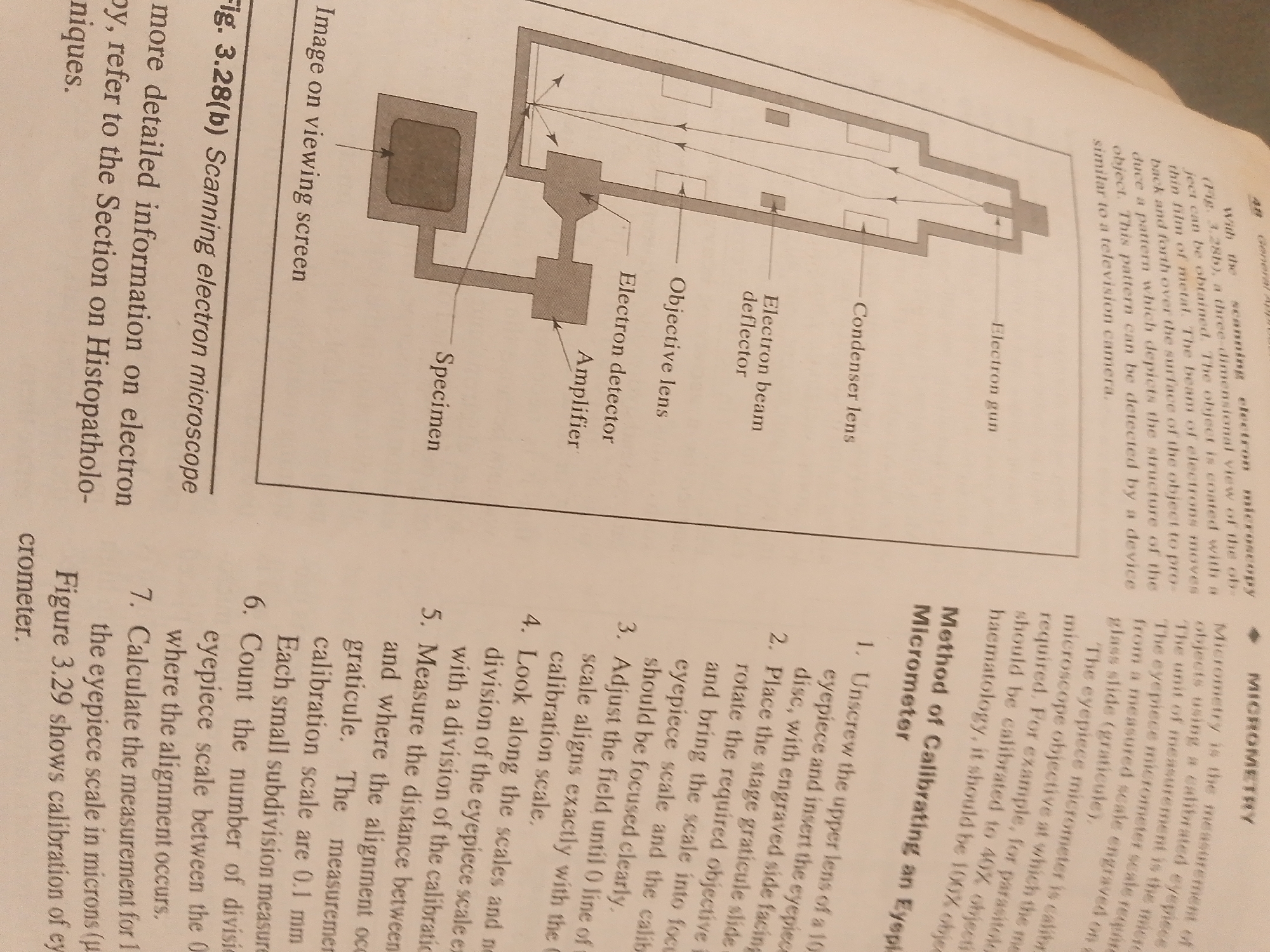
This media file contains a photograph of a page from a textbook, likely on microscopy. The page is primarily text with a diagram illustrating a scanning electron microscope. The diagram, labeled "Fig. 3.28(b) Scanning electron microscope," shows the key components of the instrument, including the electron gun, condenser lens, electron beam deflector, objective lens, electron detector, amplifier, and specimen stage. An "Image on viewing screen" is also depicted. To the left of the diagram, there is descriptive text explaining the function of a scanning electron microscope and referencing "Histopathology techniques." To the right of the diagram, there is a section titled "MICROMETRY" with a subheading "Method of Calibrating an Eyepiece Micrometer." This section provides a numbered, step-by-step procedure for calibrating a microscope's eyepiece, detailing how to use a stage graticule for accurate measurements. The text also mentions the unit of measurement being the "micrometer" and discusses calibration for different objectives like "40X objective" and "100% objective." The page is from a printed publication, indicated by the page number "48" at the top right. The lighting suggests it is an indoor photograph, likely taken with a flash or good ambient light. The overall setting is that of a study or library environment where such textbooks would be found. There are no people or explicit location cues in the image beyond the content of the textbook itself.
No transactions found