Stake attention in this memory
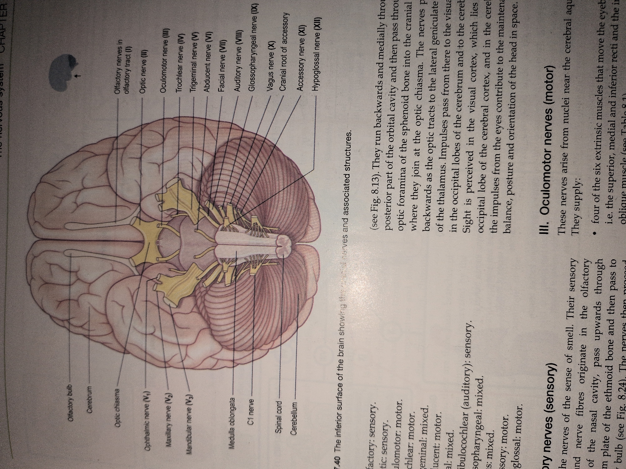
The image presents a detailed anatomical illustration of the inferior surface of the human brain, typical of a scientific or medical textbook. The central subject is a superior-inferior view of the brain, depicting the cerebrum, cerebellum, and brainstem structures. Numerous cranial nerves are distinctly labeled, originating from or projecting into the brainstem. On the left side of the illustration, structures identified include the Olfactory bulb, Cerebrum, Optic chiasma, Ophthalmic nerve (V1), Maxillary nerve (V2), Mandibular nerve (V3), Medulla oblongata, C1 nerve, Spinal cord, and Cerebellum. On the right side of the illustration, labeled structures include Olfactory nerves in olfactory tract (I), Optic nerve (II), Oculomotor nerve (III), Trochlear nerve (IV), Trigeminal nerve (V), Abducent nerve (VI), Facial nerve (VII), Auditory nerve (VIII), Glossopharyngeal nerve (IX), Vagus nerve (X), Cranial root of accessory, Accessory nerve (XI), and Hypoglossal nerve (XII). The illustration is accompanied by textual annotations below, identifying the figure as "7.40 The inferior surface of the brain showing the cranial nerves and associated structures." Further text describes the sensory and motor functions of several cranial nerves, such as "olfactory: sensory" and "oculomotor: motor," and provides detailed descriptions of "Olfactory nerves (sensory)" and "Oculomotor nerves (motor)," including their origins and supply targets. The image is a static, two-dimensional drawing on what appears to be a textbook page, indicated by chapter headers ("THE NERVOUS SYSTEM CHAPTER"), figure numbering, and descriptive text sections. There are no individuals, actions, or interactions depicted within the illustration itself, nor is there any visual information regarding a specific geographical location or environment beyond the context of a printed educational material.
Symbol
56281
Volume
5,128
Creator
+$0.00
Revenue
+$0.00
TVL
$5.00