
Sign in
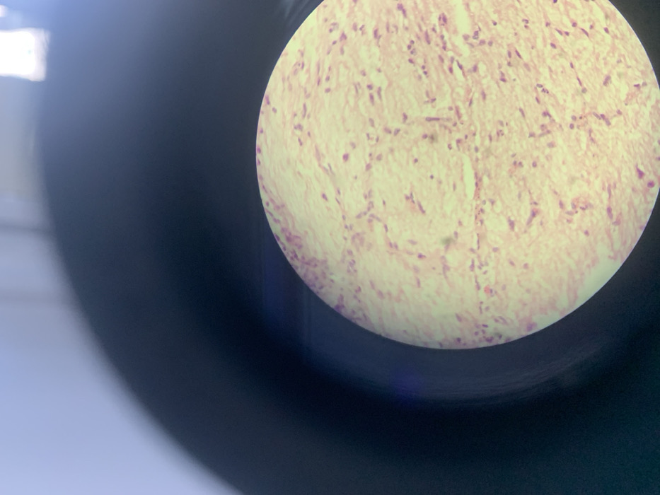
A microscopic view reveals a tissue sample, stained with a light pink hue. Scattered throughout are small, darker purple spots, likely representing cell nuclei. The tissue exhibits a fibrous, somewhat disorganized structure, possibly indicating connective tissue. The image is captured through a circular lens, with the surrounding area appearing dark and out of focus.
Emma🇨🇳💕

Dec 8, 2025, 1:25 AM

Shenyang City, China

Stake attention in this memory

microscopic image
histology
tissue
cells
pathology

A microscopic view reveals a tissue sample, stained with a light pink hue. Scattered throughout are small, darker purple spots, likely representing cell nuclei. The tissue exhibits a fibrous, somewhat disorganized structure, possibly indicating connective tissue. The image is captured through a circular lens, with the surrounding area appearing dark and out of focus.

Loading AttnAds…

transactions
revenues
stakers
Earliest
Latest
Highest stake

No transactions found

More from this user

More from Shenyang City

Emma🇨🇳💕

Dec 8, 2025, 1:25 AM

Shenyang City, China

Licensed under Creative Commons Attribution 4.0 International License. You are free to share and adapt this content with proper attribution.