
Stake attention in this memory
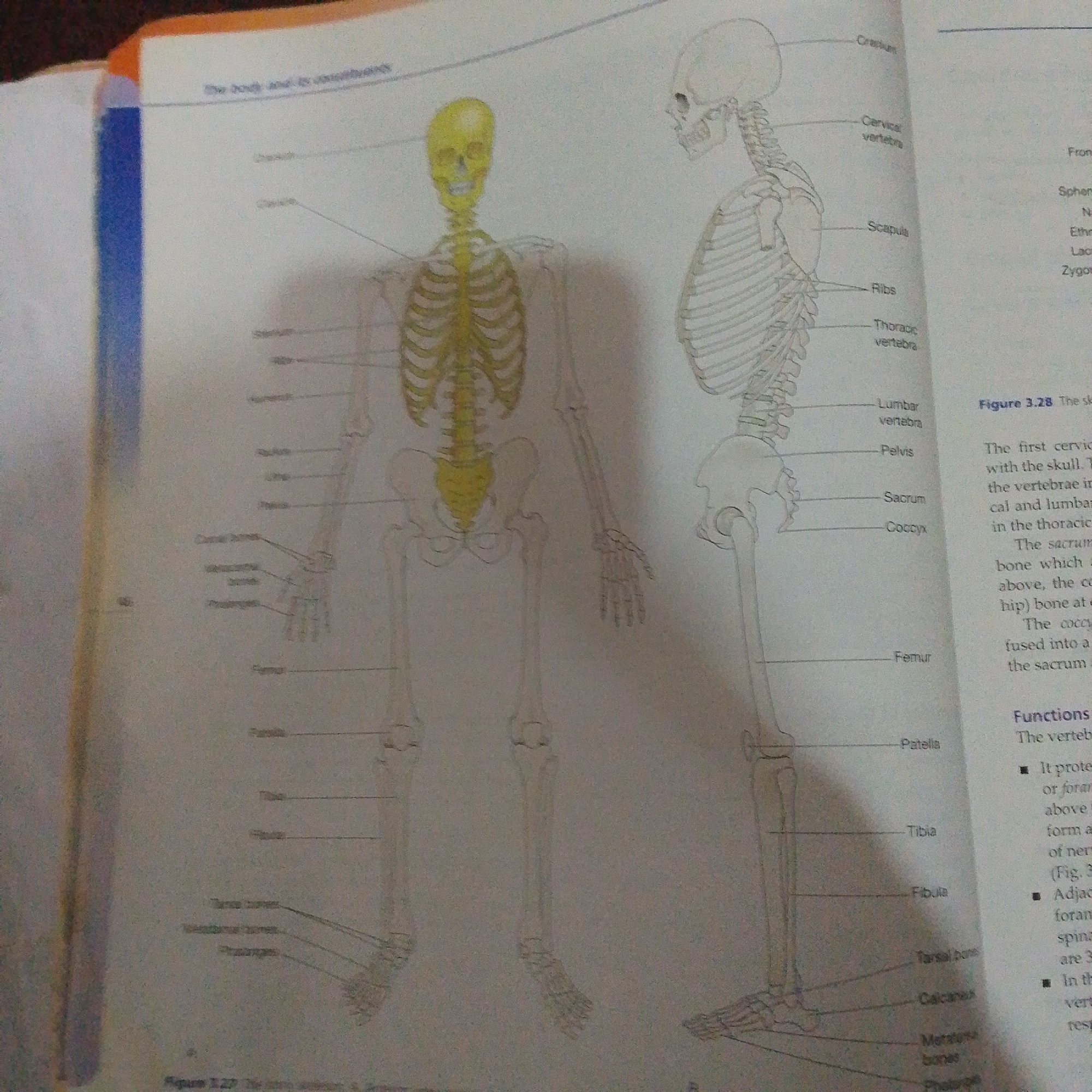
This media file is an image from a textbook, featuring anatomical diagrams of the human skeleton. The main subjects are two detailed illustrations of the human skeleton, one from the anterior view and one from the lateral view, with various parts labeled. There are no people or a specific scene depicted, only the skeletal structures. The image appears to be from a medical or biological educational context. The illustrations highlight different sections of the vertebral column (cervical, thoracic, lumbar), ribs, scapula, pelvis, sacrum, coccyx, and bones of the limbs and feet. Text labels accompany the diagrams, identifying bones like the cranium, femur, patella, tibia, fibula, tarsal bones, calcaneus, metatarsal bones, and phalanges. Visible text includes: - "The body and its components" (likely a chapter or section title) - "Cranium" - "Cervical vertebra" - "Scapula" - "Ribs" - "Thoracic vertebra" - "Lumbar vertebra" - "Pelvis" - "Sacrum" - "Coccyx" - "Femur" - "Patella" - "Tibia" - "Fibula" - "Tarsal bone" - "Calcaneus" - "Metatarsal bones" - "Phalanges" - "Figure 3.28 The sk" (partially visible) - "The first cervic with the skull. T the vertebrae in cal and lumbar in the thoracic" (part of a descriptive text) - "The sacrum bone which above, the co hip) bone at" (part of a descriptive text) - "The cocc fused into a the sacrum a" (part of a descriptive text) - "Functions" - "The verteb" - Bullet points describing protective functions of vertebrae. - "Figure 3.27 The" (partially visible) There are no cues regarding the time of day, weather, or a specific location within Nigeria. The image's context is purely educational, focusing on anatomical information. The overall tone is informative and neutral.
Loading AttnAds…
No transactions found