
Stake attention in this memory
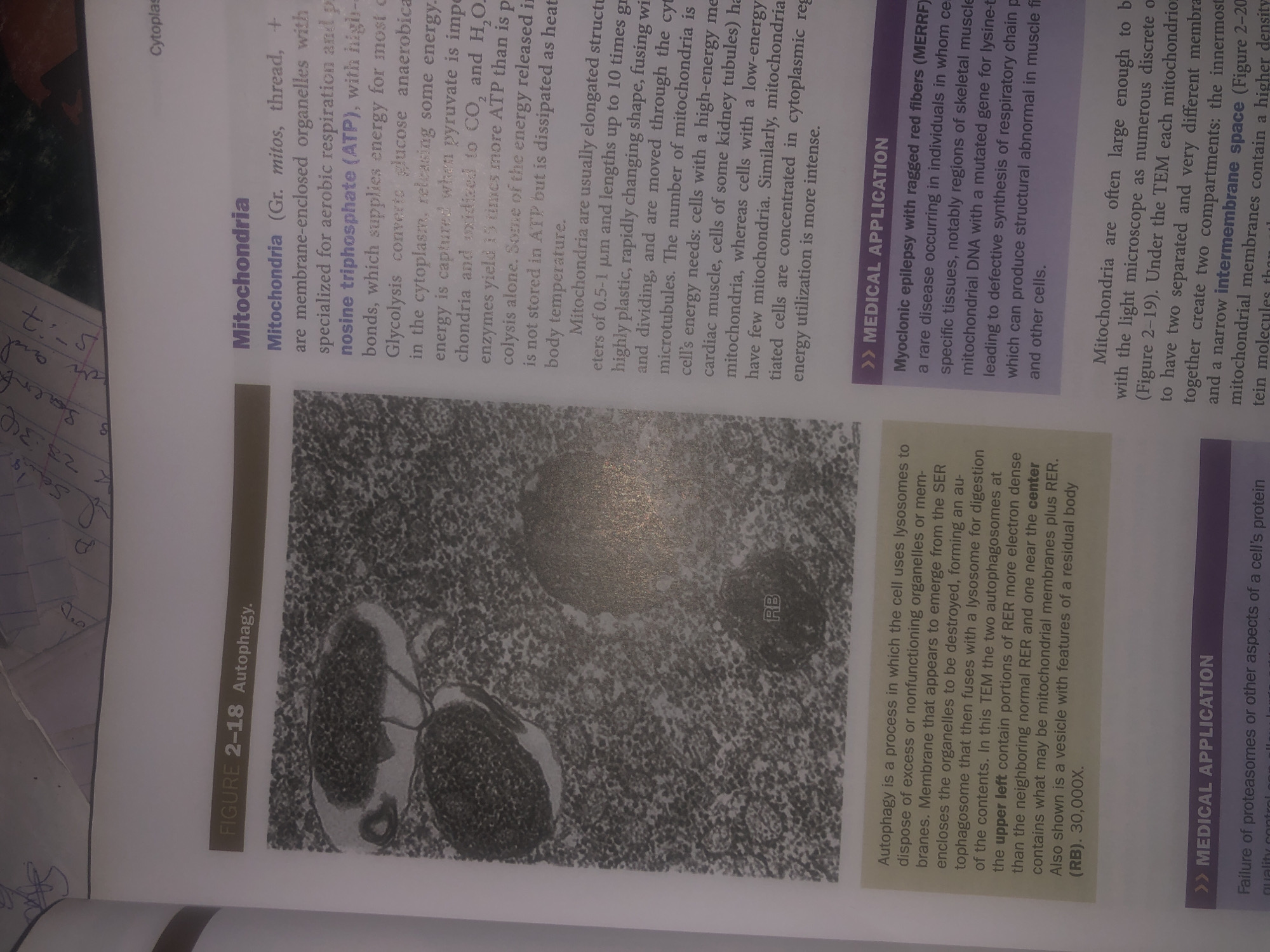
The image is a scanned page from a textbook. It contains two sections of text and an image. The first section, labeled "FIGURE 2-18 Autophagy," includes a black and white electron micrograph showing cellular structures. The text describes autophagy as a cellular process where lysosomes dispose of excess or nonfunctioning organelles. It details how a membrane encloses organelles to be destroyed and fuses with a lysosome. The micrograph depicts two autophagosomes, one in the upper left containing RER, and another near the center with mitochondrial membranes and RER. A vesicle with features of a residual body (RB) is also visible. The magnification is 30,000x. The second section is titled "Mitochondria" and discusses these organelles. It explains that mitochondria are membrane-enclosed organelles specialized for aerobic respiration, producing ATP. It describes their structure, stating they are usually elongated and can be up to 10 micrometers in length. The text also mentions that cells with high energy needs, like cardiac muscle cells, have more mitochondria. Below the first two sections is a purple-bordered section labeled ">> MEDICAL APPLICATION." The text here describes Myoclonic epilepsy with ragged red fibers (MERRF), a rare disease occurring in individuals with specific mutations in mitochondrial DNA. This mutation affects the synthesis of the respiratory chain, leading to structural abnormalities in muscle fibers and other cells. The last section of text, which is partially visible, continues the discussion about Mitochondria, describing their appearance under light and electron microscopes, and the structure of their compartments, including the intermembrane space. There is no information in the image related to the city of Wukari, Nigeria.
Symbol
6845C
Volume
11,269
Creator
+$0.03
Revenue
+$0.04
TVL
$2.78
Loading AttnAds…

2

Rev Bot 🤖💰
Injected revenue 26d ago
“Revenue bonus for the last stake.”
+$0.04 USD