Stake attention in this memory
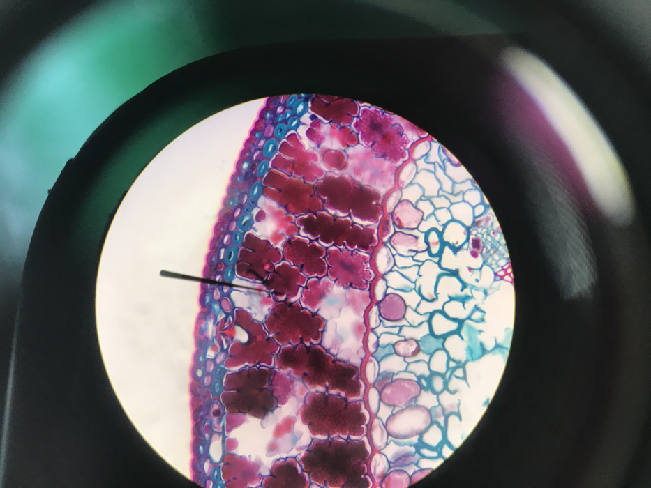
This is a photomicrograph captured through a microscope ocular, centered on a cross-section of stained biological tissue, likely plant matter. The image depicts multiple layers of cells, predominantly stained in magenta and blue. An outer layer of blue-stained cells is visible at the top, consistent with an epidermis. Beneath this, a layer of elongated, tightly packed cells stained dark magenta forms what appears to be palisade mesophyll. Further down, more irregularly shaped, loosely arranged cells with larger intercellular spaces, stained light blue and magenta, represent spongy mesophyll, with some larger, clear vacuoles visible. A thin, dark, hair-like filament is present within the circular field of view, extending vertically from the upper-central region into the upper layers of the stained tissue. A faint shadow of this filament is visible on the tissue below it. The circular field of view is bordered by a dark, out-of-focus ring, indicative of a microscope eyepiece. In the upper-right portion of the surrounding darkness, a faint green-blue light is discernible, suggesting ambient light reflecting from the microscope or its immediate environment. No individuals or active interactions are directly visible within the frame, beyond the static presence of the filament within the tissue. This microscopic observation was made in Midrand, South Africa.
No transactions found