
Stake attention in this memory
histology
textbook
salivary gland
microscopic image
education
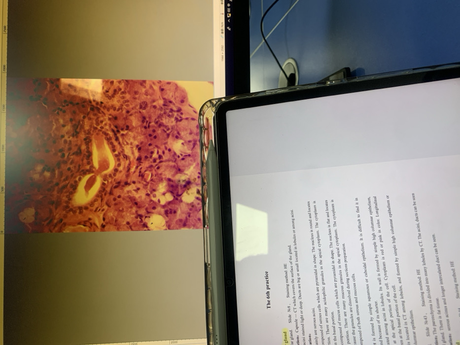
The image shows a microscopic slide of a salivary gland stained with HE. It displays serous acini, characterized by pyramidal cells, round nuclei, and acidophilic granules. Ducts are visible, appearing as either small or large structures located within lobules or among the acini. The slide also references a capsule of CT and the parenchyma divided into lobules. The accompanying text on a tablet describes the salivary gland's structure, staining methods, and key features.
Loading AttnAds…
transactions
revenues
stakers
Earliest
Latest
Highest stake
No transactions found