Sign in
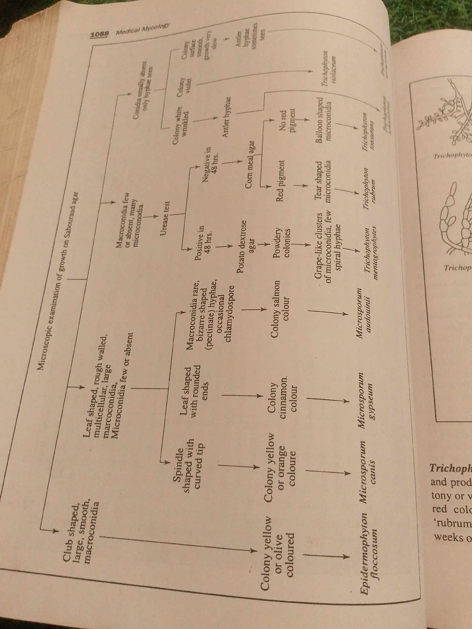
The image is a close-up of a page from a medical textbook, likely discussing medical mycology. The main subject is a diagnostic key or flowchart used to identify fungal species based on their microscopic and macroscopic characteristics.

The flowchart details examinations performed on Sabouraud agar, Corn meal agar, and Potato dextrose agar. It describes features such as the shape and presence of macroconidia and microconidia, colony color, pigment production, and urease test results. The flowchart branches out to identify different species, including various *Trichophyton* species (*T. violaceum*, *T. tonsurans*, *T. rubrum*, *T. mentagrophytes*), *Microsporum* species (*M. audouinii*, *M. canis*, *M. gypseum*), and *Epidermophyton floccosum*.

Visible text includes the chapter title "1088 Medical Mycology," descriptions of laboratory media ("Sabouraud agar," "Corn meal agar," "Potato dextrose agar"), and names of fungal genera and species. There are also labels for microscopic features like "macroconidia," "microconidia," "hyphae," and "chlamydospore." Another section on the right side of the page shows illustrations of fungal structures and begins with text referencing "Trichophyton" and "Trichophyto."

The setting is an indoor environment, likely a library or study space, as suggested by the book format and the presence of what appears to be a green fabric or surface in the background at the top of the image. There are no people, animals, or specific environmental cues like time of day or weather. The overall mood is educational and informative.
FM-qTLKq1

Jun 29, 2026, 3:35 PM

Jauro Yinu, Nigeria

Stake attention in this memory

analytical
informative
scientific
complex
detailed

The image is a close-up of a page from a medical textbook, likely discussing medical mycology. The main subject is a diagnostic key or flowchart used to identify fungal species based on their microscopic and macroscopic characteristics. The flowchart details examinations performed on Sabouraud agar, Corn meal agar, and Potato dextrose agar. It describes features such as the shape and presence of macroconidia and microconidia, colony color, pigment production, and urease test results. The flowchart branches out to identify different species, including various *Trichophyton* species (*T. violaceum*, *T. tonsurans*, *T. rubrum*, *T. mentagrophytes*), *Microsporum* species (*M. audouinii*, *M. canis*, *M. gypseum*), and *Epidermophyton floccosum*. Visible text includes the chapter title "1088 Medical Mycology," descriptions of laboratory media ("Sabouraud agar," "Corn meal agar," "Potato dextrose agar"), and names of fungal genera and species. There are also labels for microscopic features like "macroconidia," "microconidia," "hyphae," and "chlamydospore." Another section on the right side of the page shows illustrations of fungal structures and begins with text referencing "Trichophyton" and "Trichophyto." The setting is an indoor environment, likely a library or study space, as suggested by the book format and the presence of what appears to be a green fabric or surface in the background at the top of the image. There are no people, animals, or specific environmental cues like time of day or weather. The overall mood is educational and informative.

Loading AttnAds…

transactions
revenues
stakers
Earliest
Latest
Highest stake

No transactions found

More from this user

More from Jauro Yinu

FM-qTLKq1

Jun 29, 2026, 3:35 PM

Jauro Yinu, Nigeria

Licensed under Creative Commons Attribution 4.0 International License. You are free to share and adapt this content with proper attribution.