
Stake attention in this memory
X-ray
medical imaging
spine
radiographic technique
medical textbook
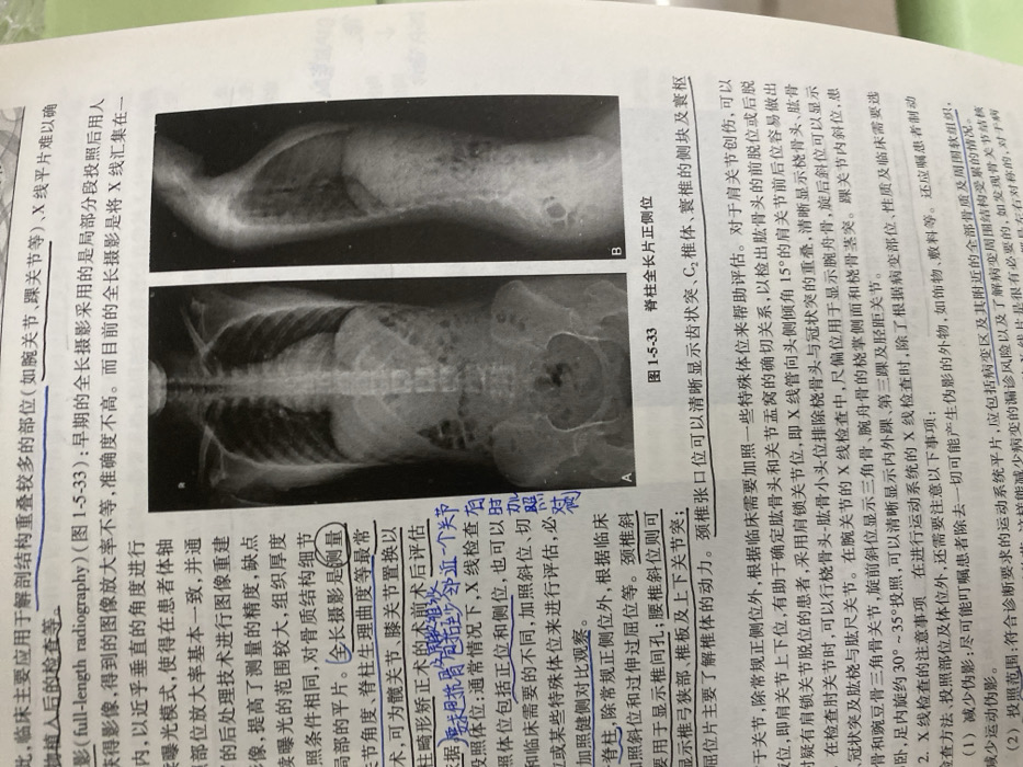
The text discusses full-length radiography used for areas with overlapping anatomy like wrists and ankles. Early methods involved segmented exposures, but current methods use a single exposure. The technique is valuable for measuring angles, spinal curvature, and pre/post-surgical evaluations. Imaging protocols mandate at least one joint beyond the area of interest. Standard positions are anterior-posterior and lateral, with variations for specific structures. The text also discusses considerations for X-ray studies of other joints like the shoulder, elbow, wrist, and ankle. Figure 1-5-33 shows anterior-posterior and lateral X-rays of the spine.
Loading AttnAds…
transactions
revenues
stakers
Earliest
Latest
Highest stake
No transactions found