Stake attention in this memory
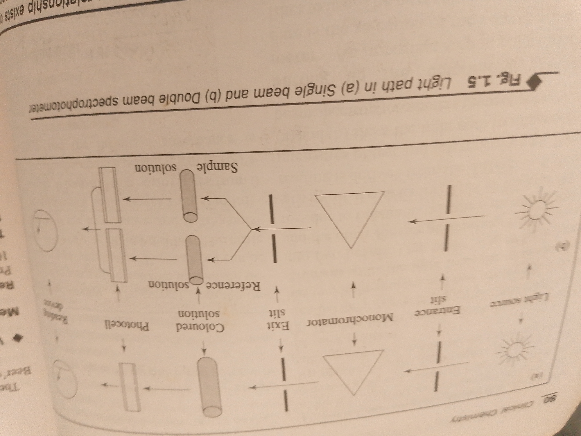
The image is a diagram illustrating the light path in single-beam and double-beam spectrophotometers, labeled as Figure 1.5. The diagram depicts various optical components involved in spectrophotometry, such as light sources, slits, monochromators, sample holders (cuvettes), photocells, and reading devices. It is presented as part of a textbook or academic material, with visible text including labels for components like "Sample solution," "Reference solution," "Monochromator," "Photocell," and "Light source." There are also section headings and page numbers, such as "Fig. 1.5," "Clinical Chemistry," and "80." The diagram is split into two parts, labeled (a) for a single-beam spectrophotometer and (b) for a double-beam spectrophotometer, indicating a comparison of their operational light paths. The overall setting appears to be an educational or scientific context. There are no people, specific locations, time of day, or weather cues visible in the image. The focus is purely on the technical illustration of the instrument's functionality.
No transactions found