
Sign in
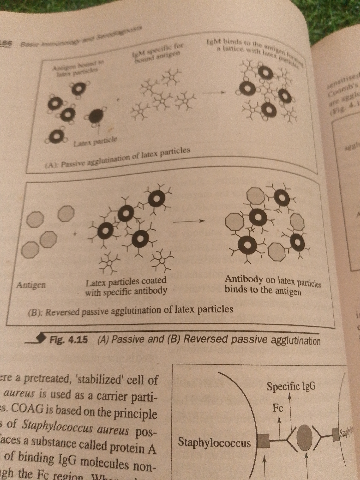
The image displays a diagram illustrating two types of agglutination reactions involving latex particles and antigens or antibodies, labeled as (A) Passive and (B) Reversed passive agglutination.

Diagram (A) shows "Antigen bound to latex particles" on the left, where small circles (latex particles) are depicted with attached antigens. These then react with "IgM specific for bound antigen" which are depicted as Y-shaped molecules, forming a lattice structure with the latex particles. The text indicates "Latex particle" and "Passive agglutination of latex particles".

Diagram (B) shows "Antigen" on the left, depicted as octagonal shapes. These are mixed with "Latex particles coated with specific antibody," where the Y-shaped antibody molecules are attached to the latex particles. The reaction results in an agglutination where the antigens bridge the antibody-coated latex particles, forming a lattice. The text indicates "Antibody on latex particles binds to the antigen" and "Reversed passive agglutination of latex particles".

Below the diagrams, there is a caption "Fig. 4.15 (A) Passive and (B) Reversed passive agglutination".

Further text to the left discusses a pre-treated, stabilized cell of *aureus* used as a carrier particle and COAG (likely Coagglutination) based on *Staphylococcus aureus* having protein A for binding IgG molecules.

To the right, a schematic diagram shows a *Staphylococcus* cell interacting with "Specific IgG" where the Fc region of the antibody is involved.

The image is a page from a textbook or scientific publication, showing diagrams related to immunology and serodiagnosis. The setting is not a physical location but a page within a book, and therefore, has no relation to Jalingo, Nigeria.
FM-qTLKq1

Jun 29, 2026, 3:28 PM

Jalingo, Nigeria

REMOVED

Stake attention in this memory

informative
scientific
educational
illustrative
diagrammatic

The image displays a diagram illustrating two types of agglutination reactions involving latex particles and antigens or antibodies, labeled as (A) Passive and (B) Reversed passive agglutination. Diagram (A) shows "Antigen bound to latex particles" on the left, where small circles (latex particles) are depicted with attached antigens. These then react with "IgM specific for bound antigen" which are depicted as Y-shaped molecules, forming a lattice structure with the latex particles. The text indicates "Latex particle" and "Passive agglutination of latex particles". Diagram (B) shows "Antigen" on the left, depicted as octagonal shapes. These are mixed with "Latex particles coated with specific antibody," where the Y-shaped antibody molecules are attached to the latex particles. The reaction results in an agglutination where the antigens bridge the antibody-coated latex particles, forming a lattice. The text indicates "Antibody on latex particles binds to the antigen" and "Reversed passive agglutination of latex particles". Below the diagrams, there is a caption "Fig. 4.15 (A) Passive and (B) Reversed passive agglutination". Further text to the left discusses a pre-treated, stabilized cell of *aureus* used as a carrier particle and COAG (likely Coagglutination) based on *Staphylococcus aureus* having protein A for binding IgG molecules. To the right, a schematic diagram shows a *Staphylococcus* cell interacting with "Specific IgG" where the Fc region of the antibody is involved. The image is a page from a textbook or scientific publication, showing diagrams related to immunology and serodiagnosis. The setting is not a physical location but a page within a book, and therefore, has no relation to Jalingo, Nigeria.

Loading AttnAds…

transactions
revenues
stakers
Earliest
Latest
Highest stake

No transactions found

More from this user

More from Jalingo

FM-qTLKq1

Jun 29, 2026, 3:28 PM

Jalingo, Nigeria

Licensed under Creative Commons Attribution 4.0 International License. You are free to share and adapt this content with proper attribution.