Stake attention in this memory
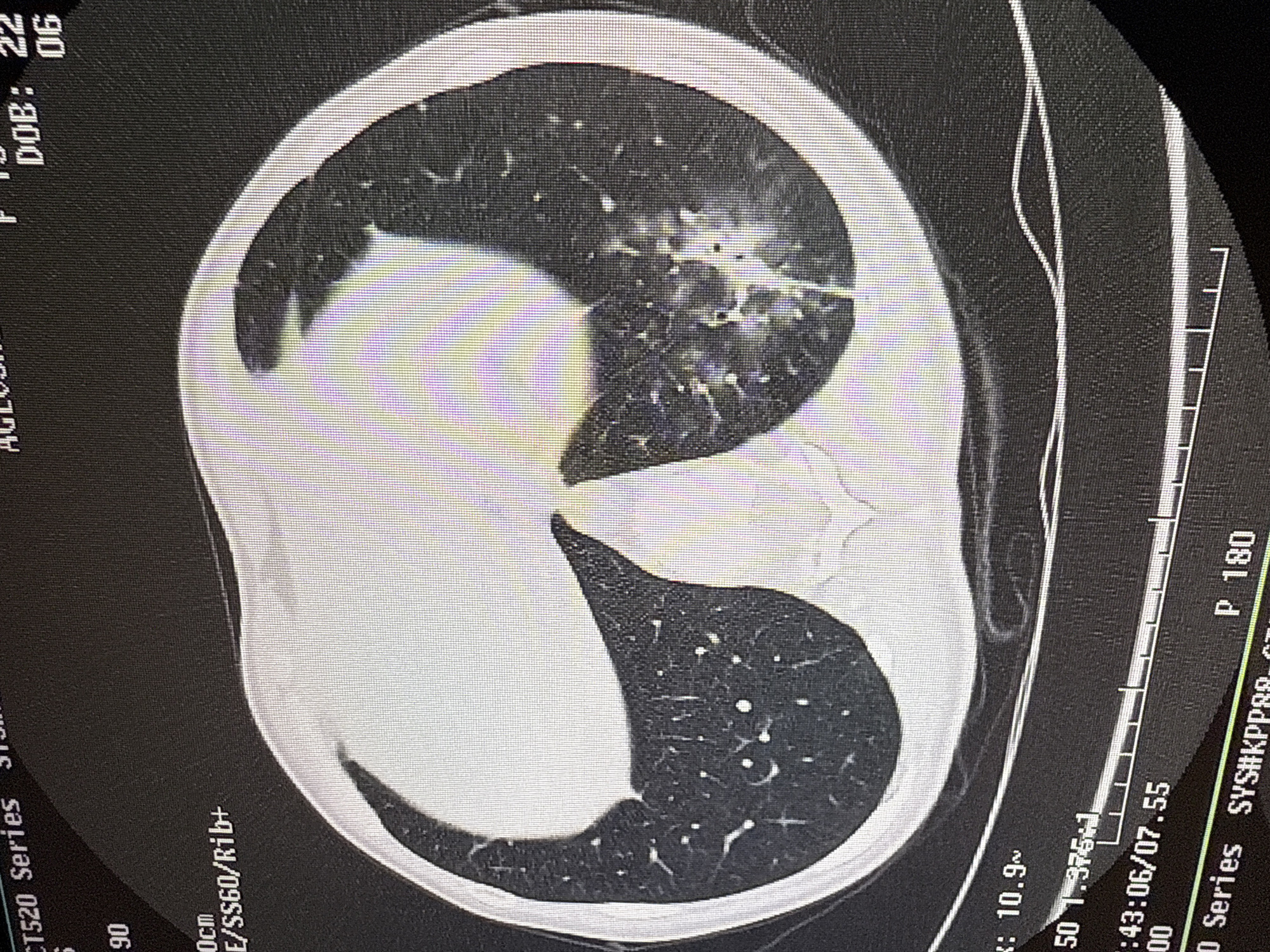
This is an axial computed tomography (CT) scan of a patient's chest, displayed on a digital screen, likely within a medical facility in Odintsovo, Russia. The central subject is the internal human anatomy, specifically the lungs and thoracic cavity, revealing significant pathology. The activity taking place is the review of a diagnostic medical image. The scan shows distinct abnormalities in both lungs. On the patient's left side (appearing on the right of the image due to standard medical imaging orientation), there is a large, dense area of consolidation or pleural effusion, largely obscuring the lung parenchyma. On the patient's right side (appearing on the left of the image), multiple patchy and nodular opacities are visible, consistent with areas of ground-glass opacification or consolidation, alongside prominent bronchial structures. The surrounding chest wall and ribs are also depicted. Notable details include the visible pixel grid of the screen display. Visible text on the image provides patient information fragments, such as "DOB: 22 06" and "AGLOBH" (partially visible), along with system identifiers like "SYS#KPP88.C". Technical parameters of the scan are also present, including "CT520 Series", "S", "90", "0cm", "E/SS60/R1b+", "P 180", "10.9~", "50 1.375:1", and a timestamp "43:06/07.55", likely indicating the time and date of the scan or image acquisition. No specific time of day or weather is directly discernible from the image itself beyond these digital timestamps.
No transactions found